Explore
 Validate
 Learn
Learn Western blot
 Immunohistochemistry
Antibody data
- Antibody Data
- Antigen structure
- References [1]
- Comments [0]
- Validations
- Immunohistochemistry [1]
Submit
Validation data
Reference
Comment
Report error
- Product number
- HPA056371 - Provider product page

- Provider
- Atlas Antibodies
- Proper citation
- Atlas Antibodies Cat#HPA056371, RRID:AB_2683108
- Product name
- Anti-FUCA1
- Antibody type
- Polyclonal
- Description
- Polyclonal Antibody against Human FUCA1, Gene description: fucosidase, alpha-L- 1, tissue, Validated applications: IHC, WB, Uniprot ID: P04066, Storage: Store at +4°C for short term storage. Long time storage is recommended at -20°C.
- Reactivity
- Human
- Host
- Rabbit
- Conjugate
- Unconjugated
- Isotype
- IgG
- Vial size
- 100 µl
- Concentration
- 0.1 mg/ml
- Storage
- Store at +4°C for short term storage. Long time storage is recommended at -20°C.
- Handling
- The antibody solution should be gently mixed before use.
Submitted references Macrophage M2 Co-expression Factors Correlate With the Immune Microenvironment and Predict Outcome of Renal Clear Cell Carcinoma
Wang Y, Yan K, Lin J, Li J, Bi J
Frontiers in Genetics 2021;12
Frontiers in Genetics 2021;12
No comments: Submit comment
Supportive validation
- Submitted by
- Atlas Antibodies (provider)
- Enhanced method
- Orthogonal validation
- Main image
- Experimental details
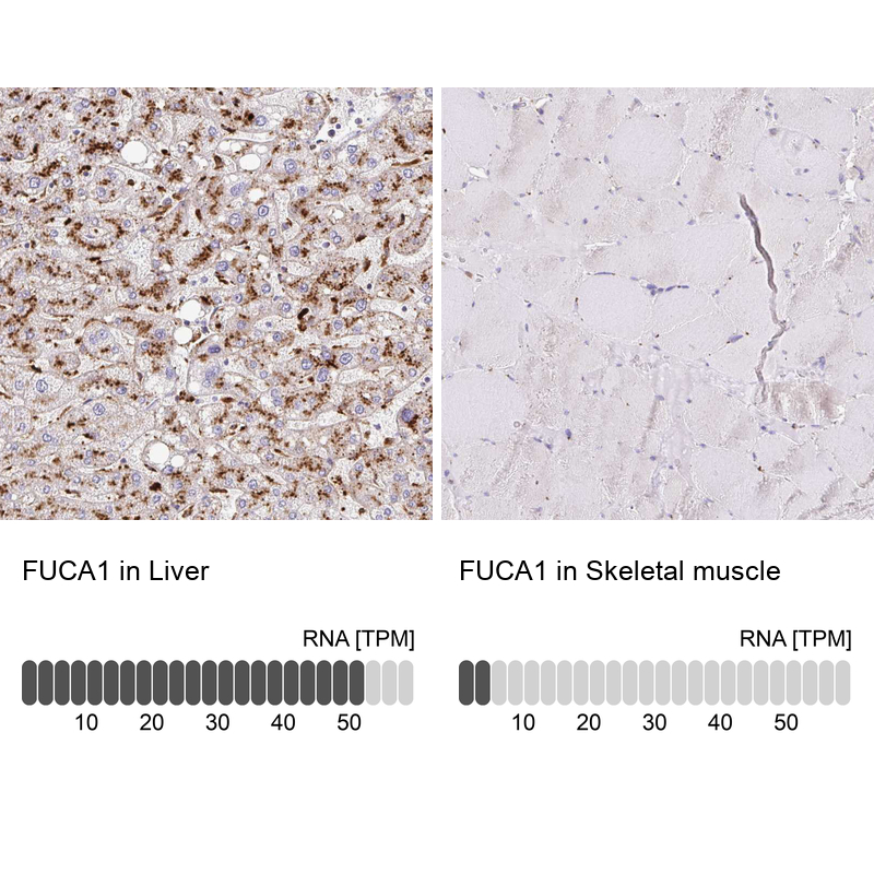
- Immunohistochemistry analysis in human liver and skeletal muscle tissues using HPA056371 antibody. Corresponding FUCA1 RNA-seq data are presented for the same tissues.
- Sample type
- Human
- Protocol
- Protocol