Explore
 Validate
 Learn
Learn Western blot
 Other assay
Antibody data
- Antibody Data
- Antigen structure
- References [2]
- Comments [0]
- Validations
- Other assay [3]
Submit
Validation data
Reference
Comment
Report error
- Product number
- PA5-44133 - Provider product page

- Provider
- Invitrogen Antibodies
- Product name
- SLC25A31 Polyclonal Antibody
- Antibody type
- Polyclonal
- Antigen
- Synthetic peptide
- Description
- Peptide sequence: LAGGVAAAVS KTAVAPIERV KLLLQVQASS KQISPEARYK GMVDCLVRIP Sequence homology: Cow: 93%; Dog: 79%; Guinea Pig: 100%; Horse: 100%; Human: 100%; Mouse: 100%; Rabbit: 93%; Rat: 100%
- Reactivity
- Human
- Host
- Rabbit
- Isotype
- IgG
- Vial size
- 100 μL
- Concentration
- 0.5 mg/mL
- Storage
- -20°C, Avoid Freeze/Thaw Cycles
Submitted references Condensed Mitochondria Assemble Into the Acrosomal Matrix During Spermiogenesis.
Extramitochondrial cardiolipin suggests a novel function of mitochondria in spermatogenesis.
Ren M, Xu Y, Phoon CKL, Erdjument-Bromage H, Neubert TA, Rajan S, Hussain MM, Schlame M
Frontiers in cell and developmental biology 2022;10:867175
Frontiers in cell and developmental biology 2022;10:867175
Extramitochondrial cardiolipin suggests a novel function of mitochondria in spermatogenesis.
Ren M, Xu Y, Erdjument-Bromage H, Donelian A, Phoon CKL, Terada N, Strathdee D, Neubert TA, Schlame M
The Journal of cell biology 2019 May 6;218(5):1491-1502
The Journal of cell biology 2019 May 6;218(5):1491-1502
No comments: Submit comment
Supportive validation
- Submitted by
- Invitrogen Antibodies (provider)
- Main image

- Experimental details
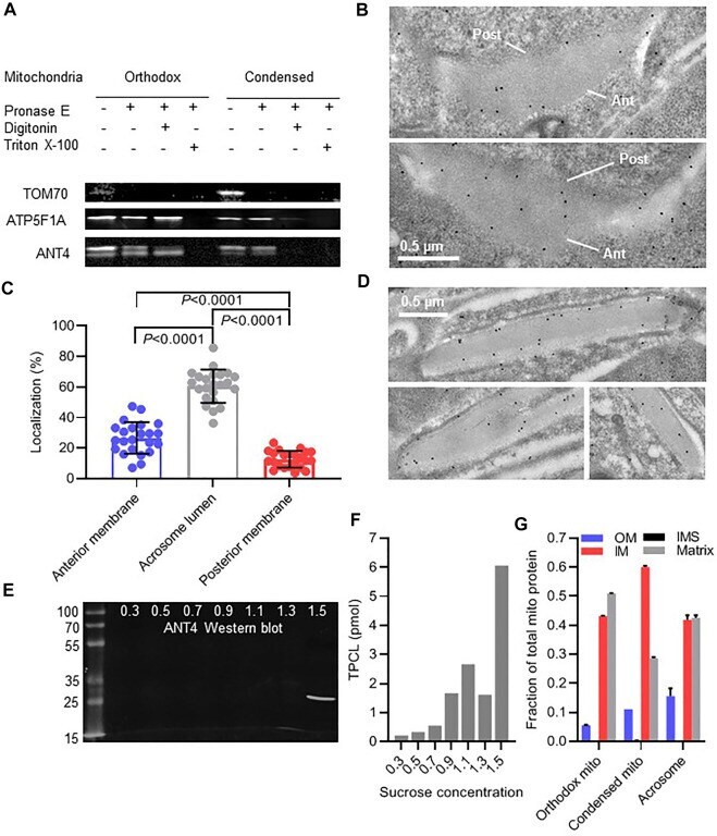
- FIGURE 5 Mitochondria are incorporated into the acrosome lumen. (A) : Isolated mitochondria from mouse testis were treated with pronase E and digitonin or Triton X-100 followed by Western blot analysis of TOM70, ATP5F1A, and ANT4. (B) : The localization of ANT4 was visualized in acrosomes of round mouse spermatids by immune electron microscopy. The anterior (Ant) and posterior (Post) membranes of the acrosome are marked. (C) : The distribution of ANT4 within acrosomes of round spermatids was determined by quantitative immune electron microscopy. Data are means +- SD of 23 cells. Groups were compared by t-test. (D) : The localization of ANT4 was visualized in acrosomes of elongated mouse spermatids by immune electron microscopy. (E,F) : Acrosomal material was released from mouse sperm and resolved on a sucrose density gradient (0.3-1.5 M sucrose). Fractions were analyzed by Western blotting (ANT4) or by mass spectrometry (TPCL). (G) : Orthodox and condensed mitochondria were purified from testis homogenate. Acrosomal material was collected from the extracellular medium of mouse sperm after inducing the acrosome reaction and further purified by sucrose density gradient centrifugation. The relative abundance of mitochondrial proteins was determined by mass spectrometry. Data show the relative abundance of the outer membrane (OM), the intermembrane space (IMS), the inner membrane (IM), and the matrix, determined by summing the signal intensities of individual proteins of these co
- Submitted by
- Invitrogen Antibodies (provider)
- Main image
- Experimental details
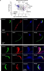
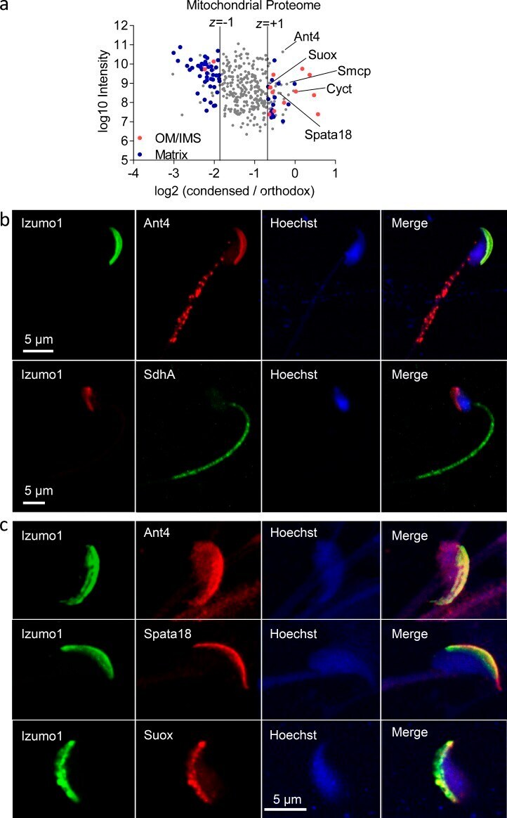
- Figure 4. The acrosome contains mitochondrial proteins. (a) Orthodox and condensed mitochondria were separated by density gradient and affinity purified. The organelles were analyzed by TMT proteomics technology. Logarithmic MS1 signal intensities were plotted against the logarithmic ratio of the MS2-based intensities of the condensed tag over the orthodox tag. Only mitochondrial proteins are shown. The data were skewed toward negative log2 ratios because mitochondrial proteins were relatively underrepresented in condensed mitochondria. Proteins with a z-score less than -1 (specific for orthodox mitochondria) and proteins with a z-score greater than +1 (specific for condensed mitochondria) were analyzed with regard to their submitochondrial localization. OM, outer membrane; IMS, intermembrane space. (b) Sperm were analyzed by immunofluorescence by using commercial antibodies to Izumo1 (acrosome marker), Ant4, and the SdhA subunit of respiratory complex II. Hoechst dye was used to stain the nucleus. (c) Sperm heads were analyzed by immunofluorescence by using antibodies to Izumo1, Ant4, Spata18, and Suox.
- Submitted by
- Invitrogen Antibodies (provider)
- Main image
- Experimental details
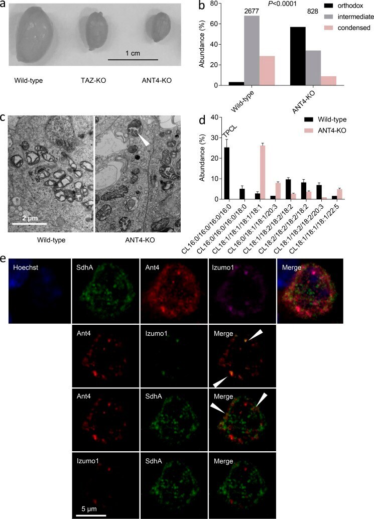
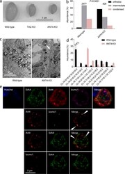
- Figure 5. Ant4 is assembled into pro-acrosomal granules. (a) Testes were excised from three mouse models (all in C57BL/6 background). (b) Orthodox, intermediate, and condensed mitochondria were quantified in all germ cells of mouse testes by using EM. The total number of mitochondria analyzed is given on top. The composition of mitochondria in wild type and ANT4-KO was compared by the chi 2 test. (c) Electron micrographs show intermediate mitochondria in mouse spermatocytes. The arrowhead points to a mitochondrion with incomplete contraction of the matrix. (d) Testis CL was analyzed by LC-MS/MS. Bar graphs show mean values +- SEMs ( n = 3). (e) Round spermatids were analyzed by immunofluorescence by using antibodies to Ant4, Izumo1 (acrosome marker), and the SdhA subunit of respiratory complex II. The nucleus was stained by Hoechst dye. The top row shows real fluorescence colors for Hoechst, SdhA, and Ant4, and magenta pseudocolor for Izumo1. Other rows show pairs of red/green pseudocolors to identify specific colocalizations. The arrowheads indicate colocalization sites.