Explore
 Validate
 Learn
Learn Immunocytochemistry
 Immunohistochemistry
Antibody data
- Antibody Data
- Antigen structure
- References [18]
- Comments [0]
- Validations
- Immunocytochemistry [1]
Submit
Validation data
Reference
Comment
Report error
- Product number
- HPA014670 - Provider product page

- Provider
- Atlas Antibodies
- Proper citation
- Atlas Antibodies Cat#HPA014670, RRID:AB_2257442
- Product name
- Anti-ZDHHC5
- Antibody type
- Polyclonal
- Description
- Polyclonal Antibody against Human ZDHHC5, Gene description: zinc finger, DHHC-type containing 5, Alternative Gene Names: KIAA1748, ZNF375, Validated applications: IHC, ICC, Uniprot ID: Q9C0B5, Storage: Store at +4°C for short term storage. Long time storage is recommended at -20°C.
- Reactivity
- Human
- Host
- Rabbit
- Conjugate
- Unconjugated
- Isotype
- IgG
- Vial size
- 100 µl
- Concentration
- 0.1 mg/ml
- Storage
- Store at +4°C for short term storage. Long time storage is recommended at -20°C.
- Handling
- The antibody solution should be gently mixed before use.
Submitted references Targeted degradation of zDHHC-PATs decreases substrate S-palmitoylation
Activity-dependent post-translational regulation of palmitoylating and depalmitoylating enzymes in the hippocampus
FASN inhibitor TVB-3166 prevents S-acylation of the spike protein of human coronaviruses
Dynamic but discordant alterations in zDHHC5 expression and palmitoylation of its substrates in cardiac pathologies
Regulation of hippocampal excitatory synapses by the Zdhhc5 palmitoyl acyltransferase
Dynamic Palmitoylation of the Sodium-Calcium Exchanger Modulates Its Structure, Affinity for Lipid-Ordered Domains, and Inhibition by XIP
Control of protein palmitoylation by regulating substrate recruitment to a zDHHC-protein acyltransferase
On the existence of endocytosis driven by membrane phase separations
CD36 facilitates fatty acid uptake by dynamic palmitoylation-regulated endocytosis
Anthrax toxin requires ZDHHC5-mediated palmitoylation of its surface-processing host enzymes
Cyclophilin A enables specific HIV-1 Tat palmitoylation and accumulation in uninfected cells
Global, site-specific analysis of neuronal protein S-acylation.
Activity-regulated trafficking of the palmitoyl-acyl transferase DHHC5
Systematic siRNA Screen Unmasks NSCLC Growth Dependence by Palmitoyltransferase DHHC5
Palmitoylation of δ-catenin by DHHC5 mediates activity-induced synapse plasticity
DHHC5 Protein Palmitoylates Flotillin-2 and Is Rapidly Degraded on Induction of Neuronal Differentiation in Cultured Cells
DHHC5 interacts with PDZ domain 3 of post-synaptic density-95 (PSD-95) protein and plays a role in learning and memory.
Zhou J, Bai M, Gallen E, Memarzadeh S, Howie J, Gao X, Kuo C, Brown E, Swingler S, Wilson S, Shattock M, France D, Fuller W
PLOS ONE 2024;19(3):e0299665
PLOS ONE 2024;19(3):e0299665
Deisl C, Moe O, Hilgemann D
2024
2024
Activity-dependent post-translational regulation of palmitoylating and depalmitoylating enzymes in the hippocampus
Abazari D, Wild A, Qiu T, Dickinson B, Bamji S
Journal of Cell Science 2023;136(7)
Journal of Cell Science 2023;136(7)
FASN inhibitor TVB-3166 prevents S-acylation of the spike protein of human coronaviruses
Mekhail K, Lee M, Sugiyama M, Astori A, St-Germain J, Latreille E, Khosraviani N, Wei K, Li Z, Rini J, Lee W, Antonescu C, Raught B, Fairn G
Journal of Lipid Research 2022;63(9):100256
Journal of Lipid Research 2022;63(9):100256
Dynamic but discordant alterations in zDHHC5 expression and palmitoylation of its substrates in cardiac pathologies
Main A, Boguslavskyi A, Howie J, Kuo C, Rankin A, Burton F, Smith G, Hajjar R, Baillie G, Campbell K, Shattock M, Fuller W
Frontiers in Physiology 2022;13
Frontiers in Physiology 2022;13
Regulation of hippocampal excitatory synapses by the Zdhhc5 palmitoyl acyltransferase
Shimell J, Globa A, Sepers M, Wild A, Matin N, Raymond L, Bamji S
Journal of Cell Science 2021;134(9)
Journal of Cell Science 2021;134(9)
Dynamic Palmitoylation of the Sodium-Calcium Exchanger Modulates Its Structure, Affinity for Lipid-Ordered Domains, and Inhibition by XIP
Gök C, Plain F, Robertson A, Howie J, Baillie G, Fraser N, Fuller W
Cell Reports 2020;31(10):107697
Cell Reports 2020;31(10):107697
Control of protein palmitoylation by regulating substrate recruitment to a zDHHC-protein acyltransferase
Plain F, Howie J, Kennedy J, Brown E, Shattock M, Fraser N, Fuller W
Communications Biology 2020;3(1)
Communications Biology 2020;3(1)
On the existence of endocytosis driven by membrane phase separations
Hilgemann D, Lin M, Fine M, Deisl C
Biochimica et Biophysica Acta (BBA) - Biomembranes 2020;1862(1):183007
Biochimica et Biophysica Acta (BBA) - Biomembranes 2020;1862(1):183007
CD36 facilitates fatty acid uptake by dynamic palmitoylation-regulated endocytosis
Hao J, Wang J, Guo H, Zhao Y, Sun H, Li Y, Lai X, Zhao N, Wang X, Xie C, Hong L, Huang X, Wang H, Li C, Liang B, Chen S, Zhao T
Nature Communications 2020;11(1)
Nature Communications 2020;11(1)
Anthrax toxin requires ZDHHC5-mediated palmitoylation of its surface-processing host enzymes
Sergeeva O, van der Goot F
Proceedings of the National Academy of Sciences 2019;116(4):1279-1288
Proceedings of the National Academy of Sciences 2019;116(4):1279-1288
Cyclophilin A enables specific HIV-1 Tat palmitoylation and accumulation in uninfected cells
Chopard C, Tong P, Tóth P, Schatz M, Yezid H, Debaisieux S, Mettling C, Gross A, Pugnière M, Tu A, Strub J, Mesnard J, Vitale N, Beaumelle B
Nature Communications 2018;9(1)
Nature Communications 2018;9(1)
Global, site-specific analysis of neuronal protein S-acylation.
Collins MO, Woodley KT, Choudhary JS
Scientific reports 2017 Jul 5;7(1):4683
Scientific reports 2017 Jul 5;7(1):4683
Activity-regulated trafficking of the palmitoyl-acyl transferase DHHC5
Brigidi G, Santyr B, Shimell J, Jovellar B, Bamji S
Nature Communications 2015;6(1)
Nature Communications 2015;6(1)
Systematic siRNA Screen Unmasks NSCLC Growth Dependence by Palmitoyltransferase DHHC5
Tian H, Lu J, Shao C, Huffman K, Carstens R, Larsen J, Girard L, Liu H, Rodriguez-Canales J, Frenkel E, Wistuba I, Minna J, Hofmann S
Molecular Cancer Research 2015;13(4):784-794
Molecular Cancer Research 2015;13(4):784-794
Palmitoylation of δ-catenin by DHHC5 mediates activity-induced synapse plasticity
Brigidi G, Sun Y, Beccano-Kelly D, Pitman K, Mobasser M, Borgland S, Milnerwood A, Bamji S
Nature Neuroscience 2014;17(4):522-532
Nature Neuroscience 2014;17(4):522-532
DHHC5 Protein Palmitoylates Flotillin-2 and Is Rapidly Degraded on Induction of Neuronal Differentiation in Cultured Cells
Li Y, Martin B, Cravatt B, Hofmann S
Journal of Biological Chemistry 2012;287(1):523-530
Journal of Biological Chemistry 2012;287(1):523-530
DHHC5 interacts with PDZ domain 3 of post-synaptic density-95 (PSD-95) protein and plays a role in learning and memory.
Li Y, Hu J, Höfer K, Wong AM, Cooper JD, Birnbaum SG, Hammer RE, Hofmann SL
The Journal of biological chemistry 2010 Apr 23;285(17):13022-31
The Journal of biological chemistry 2010 Apr 23;285(17):13022-31
No comments: Submit comment
Supportive validation
- Submitted by
- Atlas Antibodies (provider)
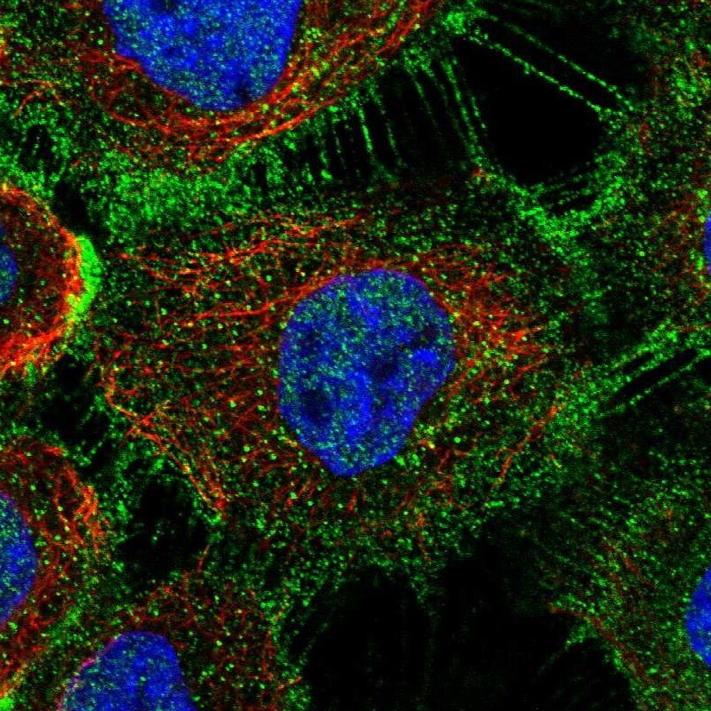
- Main image

- Experimental details
- Immunofluorescent staining of human cell line A-431 shows localization to nucleoplasm, plasma membrane & cell junctions.
- Sample type
- Human