Explore
 Validate
 Learn
Learn Western blot
 Immunohistochemistry
Antibody data
- Antibody Data
- Antigen structure
- References [0]
- Comments [0]
- Validations
- Immunohistochemistry [3]
Submit
Validation data
Reference
Comment
Report error
- Product number
- PA5-37982 - Provider product page

- Provider
- Invitrogen Antibodies
- Product name
- ZNRF1 Polyclonal Antibody
- Antibody type
- Polyclonal
- Antigen
- Synthetic peptide
- Description
- This antibody is predicted to react with human, rat, dog, pig and cow based on sequence homology. This antibody is tested in Peptide ELISA: antibody detection limit dilution 32,000.
- Reactivity
- Human, Mouse
- Host
- Goat
- Isotype
- IgG
- Vial size
- 100 μg
- Concentration
- 0.5 mg/mL
- Storage
- -20°C, Avoid Freeze/Thaw Cycles
No comments: Submit comment
Supportive validation
- Submitted by
- Invitrogen Antibodies (provider)
- Main image
- Experimental details
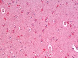
- Immunohistochemistry analysis of ZNRF1 in Human Cortex (Formalin-Fixed, Paraffin-Embedded). Samples were incubated with ZNRF1 polyclonal antibody (Product # PA5-37982).
- Submitted by
- Invitrogen Antibodies (provider)
- Main image
- Experimental details
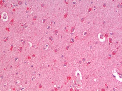
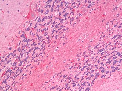
- Immunohistochemistry analysis of ZNRF1 in Human Cerebellum (Formalin-Fixed, Paraffin-Embedded). Samples were incubated with ZNRF1 polyclonal antibody (Product # PA5-37982).
- Submitted by
- Invitrogen Antibodies (provider)
- Main image
- Experimental details
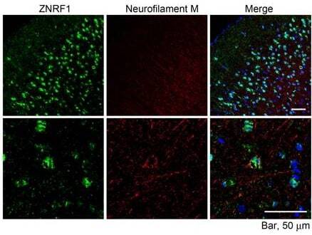
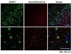
- Immunohistochemical analysis of ZNRF1 in paraffin embedded mouse cerebral cortex using a ZNRF1 polyclonal antibody (Product #PA5-37982) at a concentration of 1:100. Microwaved antigen retrieval was performed with pH 6 buffered citrate. Samples were then stained with streptavidfine-Alexa 488 after biotinylation with an anti-goat secondary antibody. The Neurofilament M was labeled with a different antibody.