Explore
 Validate
 Learn
Learn Western blot
 Immunoprecipitation
Antibody data
- Antibody Data
- Antigen structure
- References [1]
- Comments [0]
- Validations
- Immunoprecipitation [1]
- Other assay [4]
Submit
Validation data
Reference
Comment
Report error
- Product number
- PA5-81200 - Provider product page

- Provider
- Invitrogen Antibodies
- Product name
- HOXB8 Polyclonal Antibody
- Antibody type
- Polyclonal
- Antigen
- Synthetic peptide
- Description
- This product is preservative free. It is recommended to add sodium azide to avoid contamination (final concentration 0.05%-0.1%). This antibody has specificity for Human HOXB8.
- Reactivity
- Human
- Host
- Rabbit
- Isotype
- IgG
- Vial size
- 100 μL
- Concentration
- 5 mg/mL
- Storage
- Store at 4°C short term. For long term storage, store at -20°C, avoiding freeze/thaw cycles.
Submitted references Downregulation of long non-coding RNA MAFG-AS1 represses tumorigenesis of colorectal cancer cells through the microRNA-149-3p-dependent inhibition of HOXB8.
Ruan Z, Deng H, Liang M, Xu Z, Lai M, Ren H, Deng X, Su X
Cancer cell international 2020;20:511
Cancer cell international 2020;20:511
No comments: Submit comment
Supportive validation
- Submitted by
- Invitrogen Antibodies (provider)
- Main image
- Experimental details
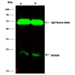
- HOXB8 Immunoprecipitation using: Lane A: 0.5 mg HepG2 Whole Cell Lysate, Lane B: 0.5 mg 293T Whole Cell Lysate 0.5 µL with HOXB8 Polyclonal Antibody (Product # PA5-81200) and 60 μg of Immunomagnetic beads Protein G. Primary antibody: HOXB8 Polyclonal Antibody, at 1:500 dilution. Secondary antibody: Dylight 800-labeled antibody to rabbit IgG (H+L), at 1:5,000 dilution. Developed using the Odyssey technique. Performed under reducing conditions. Predicted band size: 28 kDa. Observed band size: 24 kDa.

Supportive validation
- Submitted by
- Invitrogen Antibodies (provider)
- Main image

- Experimental details
- HOXB8 Immunoprecipitation using: Lane A: 0.5 mg HepG2 Whole Cell Lysate, Lane B: 0.5 mg 293T Whole Cell Lysate 0.5 µL with HOXB8 Polyclonal Antibody (Product # PA5-81200) and 60 μg of Immunomagnetic beads Protein G. Primary antibody: HOXB8 Polyclonal Antibody, at 1:500 dilution. Secondary antibody: Dylight 800-labeled antibody to rabbit IgG (H+L), at 1:5,000 dilution. Developed using the Odyssey technique. Performed under reducing conditions. Predicted band size: 28 kDa. Observed band size: 24 kDa.

- Submitted by
- Invitrogen Antibodies (provider)
- Main image

- Experimental details
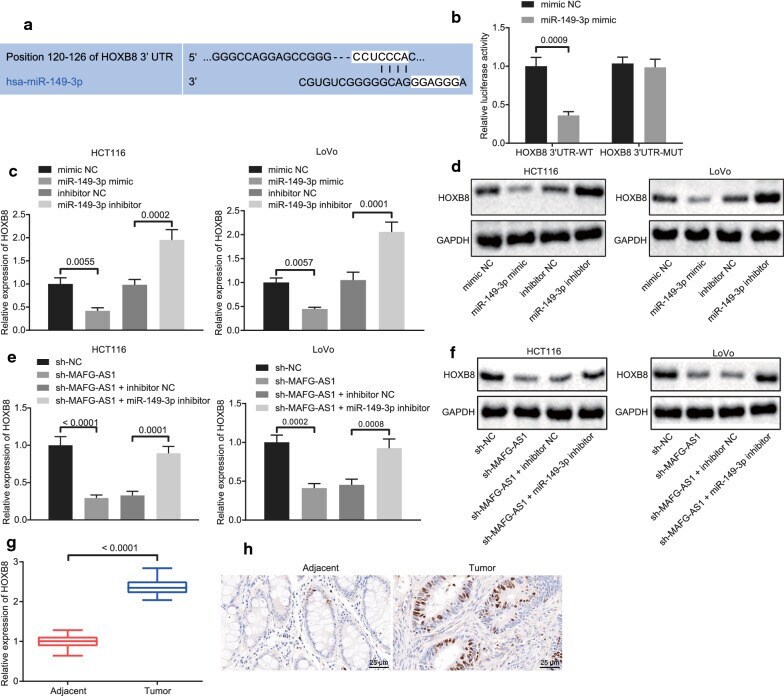
- Fig. 4 LncRNA MAFG-AS1 regulates the expression HOXB8 by targeting miR-149-3p. a The binding sites between miR-149-3p and HOXB8 predicted by Targetscan, which is an online prediction tool for biological targets of miRNA by identifying conservative 8mer, 7mer and 6mer sites that match the seed region of each miRNA. b The binding between miR-149-3p and HOXB8 detected by dual luciferase reporter gene assay. c The mRNA expression of HOXB8 in response to different expression patterns of miR-149-3p in HCT116 and LoVo cells determined by RT-qPCR. d The protein expression of HOXB8 in response to different expression patterns of miR-149-3p normalized to GAPDH in HCT116 and LoVo cells determined by Western blot analysis. e The mRNA expression of HOXB8 in response to different expression patterns of lncRNA MAFG-AS1 and miR-149-3p in HCT116 and LoVo cells determined by RT-qPCR. f The protein expression of HOXB8 in response to different expression patterns of lncRNA MAFG-AS1 and miR-149-3p normalized to GAPDH in HCT116 and LoVo cells determined by Western blot analysis. g The expression of HOXB8 in CRC tissues and adjacent tissues determined by RT-qPCR (n = 30). h The expression of HOXB8 in CRC tissues and adjacent tissues detected by immunohistochemistry (scale bar = 25 um). Data between two groups were compared by unpaired t test but by paired t test between CRC tissues and adjacent tissues. Data among multiple groups were compared by one-way ANOVA, followed by Tukey's post hoc test. Th
- Submitted by
- Invitrogen Antibodies (provider)
- Main image
- Experimental details
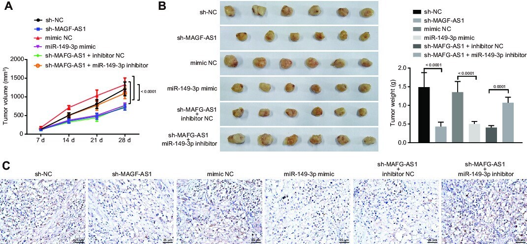
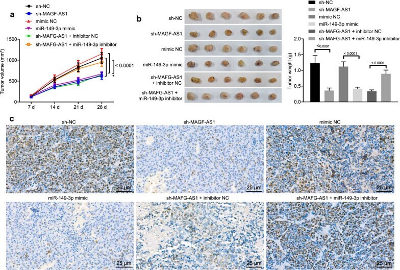
- Fig. 6 The lncRNA MAFG-AS1/miR-149-3p/HOXB8 axis promotes tumorigenesis of CRC cells (HCT116 cells) in vivo. a Tumor volume from the 7th day after injection. b Representative pictures of resected tumors and tumor weight. c The expression of HOXB8 in tumors identified by immunohistochemistry (scale bar = 25 um). Data among multiple groups were compared by one-way ANOVA, followed by Tukey's post hoc test. Data at different time points were analyzed by repeated measures ANOVA, followed by Bonferroni test. The experiment was repeated 3 times independently
- Submitted by
- Invitrogen Antibodies (provider)
- Main image
- Experimental details
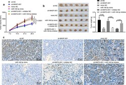
- Additional file 1: Figure S1. The lncRNA MAFG-AS1/miR-149-3p/HOXB8 axis promotes tumorigenesis of CRC cells (LoVo cells) in vivo. A, Tumor volume from the 7th day after injection. B, Representative pictures of resected tumors and tumor weight. C, The expression of HOXB8 in tumors identified by immunohistochemistry (scale bar = 25 um). Data among multiple groups were compared by one-way ANOVA, followed by Tukey's post hoc test. Data at different time points were analyzed by repeated measures ANOVA, followed by Bonferroni test. The experiment was repeated 3 times independently.