Explore
 Validate
 Learn
Learn Western blot
 Immunoprecipitation
Antibody data
- Antibody Data
- Antigen structure
- References [1]
- Comments [0]
- Validations
- Immunoprecipitation [2]
- Immunohistochemistry [5]
Submit
Validation data
Reference
Comment
Report error
- Product number
- NB600-274 - Provider product page

- Provider
- Novus Biologicals
- Proper citation
- Novus Cat#NB600-274, RRID:AB_2251999
- Product name
- Rabbit Polyclonal PAF1 Antibody
- Antibody type
- Polyclonal
- Description
- Immunogen affinity purified.
- Reactivity
- Human, Mouse
- Host
- Rabbit
- Isotype
- IgG
- Vial size
- 100 ul
- Concentration
- 1.0 mg/ml
- Storage
- Store at 4C. Do not freeze.
Submitted references The ubiquitin hydrolase USP22 contributes to 3'-end processing of JAK-STAT-inducible genes.
Chipumuro E, Henriksen MA
FASEB journal : official publication of the Federation of American Societies for Experimental Biology 2012 Feb;26(2):842-54
FASEB journal : official publication of the Federation of American Societies for Experimental Biology 2012 Feb;26(2):842-54
No comments: Submit comment
Supportive validation
- Submitted by
- Novus Biologicals (provider)
- Main image

- Experimental details
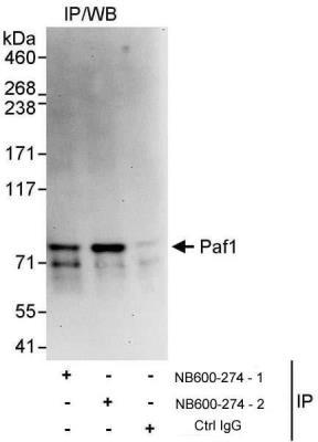
- Immunoprecipitation: PAF1 Antibody [NB600-274] - Samples: Whole cell lysate (1 mg for IP, 20% of IP loaded) from HeLa cells. Antibodies: Affinity purified rabbit anti-Paf1 antibody NB600-274 Lot 2 used for IP at 3 ug/mg lysate. Paf1 was also immunoprecipitated by rabbit anti-Paf1 Lot 1 of the same product. For blotting immunoprecipitated Paf1, NB600-274 Lot 2 was used at 1 ug/ml. Detection: Chemiluminescence with an exposure time 30 seconds.
- Submitted by
- Novus Biologicals (provider)
- Main image
- Experimental details
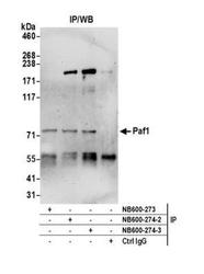
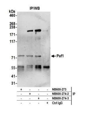
- Immunoprecipitation: PAF1 Antibody [NB600-274] - Detection of human Paf1 by western blot of immunoprecipitates. Samples: Whole cell lysate (0.5 or 1.0 mg per IP reaction; 20% of IP loaded) from HeLa cells prepared using NETN lysis buffer. Antibodies: Affinity purified rabbit anti-Paf1 antibody NB600-274 (lot NB600-274-3) used for IP at 6 ug per reaction. Paf1 was also immunoprecipitated by a previous lot of this antibody (lot NB600-274-2) and rabbit anti-Paf1 antibody NB600-273. For blotting immunoprecipitated Paf1, NB600-274 was used at 0.1 ug/ml. Detection: Chemiluminescence with an exposure time of 3 minutes.
Supportive validation
- Submitted by
- Novus Biologicals (provider)
- Main image
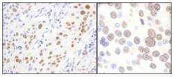
- Experimental details
- Immunohistochemistry-Paraffin: PAF1 Antibody [NB600-274] - Sections of human lung carcinoma (left) and mouse renal cell carcinoma (right).
- Submitted by
- Novus Biologicals (provider)
- Main image
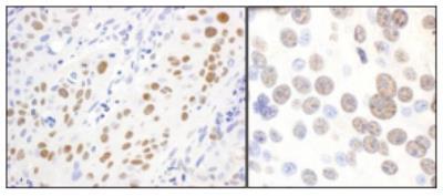
- Experimental details
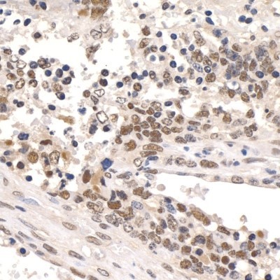
- Immunohistochemistry-Paraffin: PAF1 Antibody [NB600-274] - FFPE section of human pancreatic islet cell tumor. Affinity purified rabbit anti-Paf1 used at a dilution of 1:250.
- Submitted by
- Novus Biologicals (provider)
- Main image
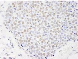
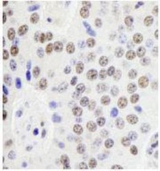
- Experimental details
- Immunohistochemistry-Paraffin: PAF1 Antibody [NB600-274] - FFPE section of human small cell lung cancer. Affinity purified rabbit anti-Paf1 used at a dilution of 1:250.
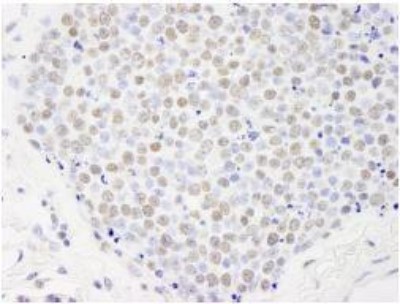
- Submitted by
- Novus Biologicals (provider)
- Main image
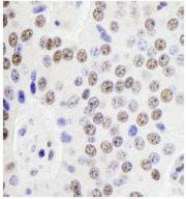
- Experimental details
- Immunohistochemistry-Paraffin: PAF1 Antibody [NB600-274] - Section of human breast carcinoma. Antibody: Affinity purified rabbit anti- Paf1 used at a dilution of 1:1,000 (1ug/ml). Detection: DAB

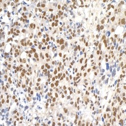
- Submitted by
- Novus Biologicals (provider)
- Main image

- Experimental details
- Immunohistochemistry-Paraffin: PAF1 Antibody [NB600-274] - Section of mouse teratoma. Antibody: Affinity purified rabbit anti- Paf1 used at a dilution of 1:1,000 (1ug/ml). Detection: DAB