Explore
 Validate
 Learn
Learn Western blot
Antibody data
- Antibody Data
- Antigen structure
- References [5]
- Comments [0]
- Validations
- Western blot [3]
- Immunoprecipitation [1]
Submit
Validation data
Reference
Comment
Report error
- Product number
- NB600-273 - Provider product page

- Provider
- Novus Biologicals
- Proper citation
- Novus Cat#NB600-273, RRID:AB_2159770
- Product name
- Rabbit Polyclonal PAF1 Antibody
- Antibody type
- Polyclonal
- Description
- Immunogen affinity purified.
- Reactivity
- Human, Mouse
- Host
- Rabbit
- Isotype
- IgG
- Vial size
- 100 ul
- Concentration
- 1.0 mg/ml
- Storage
- Store at 4C. Do not freeze.
Submitted references The PAF1 complex differentially regulates cardiomyocyte specification.
SHIP2, a factor associated with diet-induced obesity and insulin sensitivity, attenuates FGF signaling in vivo.
Defective nucleolar localization and dominant interfering properties of a parafibromin L95P missense mutant causing the hyperparathyroidism-jaw tumor syndrome.
hCTR9, a component of Paf1 complex, participates in the transcription of interleukin 6-responsive genes through regulation of STAT3-DNA interactions.
Nuclear localization of the parafibromin tumor suppressor protein implicated in the hyperparathyroidism-jaw tumor syndrome enhances its proapoptotic function.
Langenbacher AD, Nguyen CT, Cavanaugh AM, Huang J, Lu F, Chen JN
Developmental biology 2011 May 1;353(1):19-28
Developmental biology 2011 May 1;353(1):19-28
SHIP2, a factor associated with diet-induced obesity and insulin sensitivity, attenuates FGF signaling in vivo.
Jurynec MJ, Grunwald DJ
Disease models & mechanisms 2010 Nov-Dec;3(11-12):733-42
Disease models & mechanisms 2010 Nov-Dec;3(11-12):733-42
Defective nucleolar localization and dominant interfering properties of a parafibromin L95P missense mutant causing the hyperparathyroidism-jaw tumor syndrome.
Panicker LM, Zhang JH, Dagur PK, Gastinger MJ, Simonds WF
Endocrine-related cancer 2010 Jun;17(2):513-24
Endocrine-related cancer 2010 Jun;17(2):513-24
hCTR9, a component of Paf1 complex, participates in the transcription of interleukin 6-responsive genes through regulation of STAT3-DNA interactions.
Youn MY, Yoo HS, Kim MJ, Hwang SY, Choi Y, Desiderio SV, Yoo JY
The Journal of biological chemistry 2007 Nov 30;282(48):34727-34
The Journal of biological chemistry 2007 Nov 30;282(48):34727-34
Nuclear localization of the parafibromin tumor suppressor protein implicated in the hyperparathyroidism-jaw tumor syndrome enhances its proapoptotic function.
Lin L, Czapiga M, Nini L, Zhang JH, Simonds WF
Molecular cancer research : MCR 2007 Feb;5(2):183-93
Molecular cancer research : MCR 2007 Feb;5(2):183-93
No comments: Submit comment
Supportive validation
- Submitted by
- Novus Biologicals (provider)
- Main image
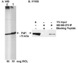
- Experimental details
- Western Blot: PAF1 Antibody [NB600-273] - Detection of Human Paf1 on HeLa whole cell lysate using NB600-273. In B, Paf1 was immunoprecipitated using NB600-274 and blotted using NB600-273 at 0.2 mcg/ml.

- Submitted by
- Novus Biologicals (provider)
- Main image
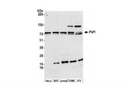
- Experimental details
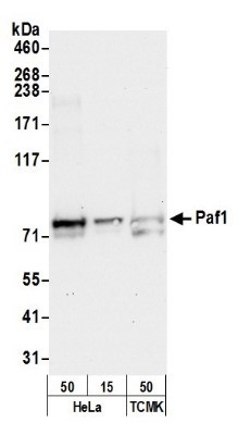
- Western Blot: PAF1 Antibody [NB600-273] - Whole cell lysate (15 ug) from HeLa, 293T, Jurkat, mouse TCMK-1, and mouse NIH3T3 cells prepared using NETN lysis buffer. Antibody: Affinity purified rabbit antiPaf1 antibody used for WB at 0.1 ug/ml. Detection: Chemiluminescence with an exposure time of 3 minutes.
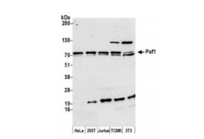
- Submitted by
- Novus Biologicals (provider)
- Main image
- Experimental details
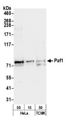
- Western Blot: PAF1 Antibody [NB600-273] - Detection of Human and Mouse Paf1 by Western Blot. Samples: Whole cell lysate (50, 15 ug) from HeLa and (50 ug) from Mouse TCMK-1 cells prepared using NETN lysis buffer. Antibody: Affinity purified rabbit anti-Paf1 antibody NB600-273 used for WB at 0.4 ug/ml. Detection: Chemiluminescence with an exposure time of 30 seconds.
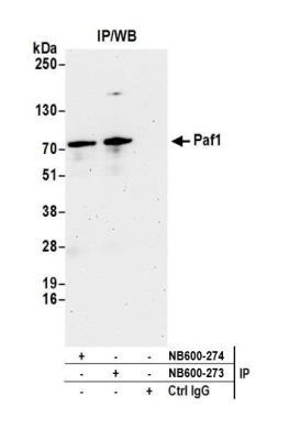
Supportive validation
- Submitted by
- Novus Biologicals (provider)
- Main image
- Experimental details
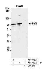
- Immunoprecipitation: PAF1 Antibody [NB600-273] - Detection of human Paf1 by western blot of immunoprecipitates. Samples: Whole cell lysate (1.0 mg per IP reaction; 20% of IP loaded) from HeLa cells prepared using NETN lysis buffer. Antibodies: Affinity purified rabbit anti-Paf1 antibody NB600-273 used for IP at 3 ug per reaction. Paf1 was also immunoprecipitated by rabbit anti-Paf1 antibody NB600-274. For blotting immunoprecipitated Paf1, NB600-273 was used at 0.4 ug/ml. Detection: Chemiluminescence with an exposure time of 3 minutes.