 Explore
 Validate
 Learn
Learn Western blot
Antibody data
- Antibody Data
- Antigen structure
- References [1]
- Comments [0]
- Validations
- Western blot [1]
- Immunohistochemistry [1]
Submit
Validation data
Reference
Comment
Report error
- Product number
- AF6648 - Provider product page

- Provider
- Novus Biologicals
- Product name
- Sheep Polyclonal Ly6K Antibody
- Antibody type
- Polyclonal
- Description
- Immunogen affinity purified. Detects human Ly6K in Western blots.
- Reactivity
- Human
- Host
- Sheep
- Isotype
- IgG
- Vial size
- 100 ug
- Concentration
- LYOPH
- Storage
- Use a manual defrost freezer and avoid repeated freeze-thaw cycles. 12 months from date of receipt, -20 to -70 degreesC as supplied. 1 month, 2 to 8 degreesC under sterile conditions after reconstitution. 6 months, -20 to -70 degreesC under sterile conditions after reconstitution.
Submitted references Ly6E/K Signaling to TGFβ Promotes Breast Cancer Progression, Immune Escape, and Drug Resistance.
AlHossiny M, Luo L, Frazier WR, Steiner N, Gusev Y, Kallakury B, Glasgow E, Creswell K, Madhavan S, Kumar R, Upadhyay G
Cancer research 2016 Jun 1;76(11):3376-86
Cancer research 2016 Jun 1;76(11):3376-86
No comments: Submit comment
Supportive validation
- Submitted by
- Novus Biologicals (provider)
- Main image
- Experimental details
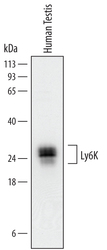
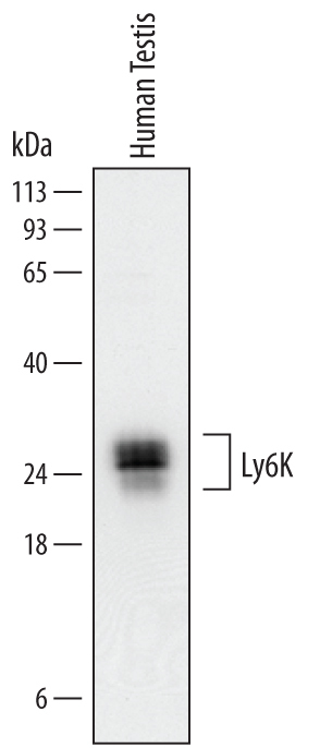
- Detection of Human Ly6K by Western Blot. Western blot shows lysates of human testis tissue. PVDF Membrane was probed with 0.25 µg/mL of Sheep Anti-Human Ly6K Antigen Affinity-purified Polyclonal Antibody (Catalog # AF6648) followed by HRP-conjugated Anti-Sheep IgG Secondary Antibody (Catalog # HAF016). Specific bands were detected for Ly6K at approximately 25-27 kDa (as indicated). This experiment was conducted under reducing conditions and using Immunoblot Buffer Group 1.
Supportive validation
- Submitted by
- Novus Biologicals (provider)
- Main image
- Experimental details
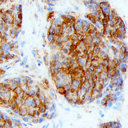
- Ly6K in Human Lung Cancer Tissue. Ly6K was detected in immersion fixed paraffin-embedded sections of human lung cancer tissue using Sheep Anti-Human Ly6K Antigen Affinity-purified Polyclonal Antibody (Catalog # AF6648) at 3 µg/mL overnight at 4 °C. Before incubation with the primary antibody, tissue was subjected to heat-induced epitope retrieval using Antigen Retrieval Reagent-Basic (Catalog # CTS013). Tissue was stained using the Anti-Sheep HRP-DAB Cell & Tissue Staining Kit (brown; Catalog # CTS019) and counterstained with hematoxylin (blue). Specific staining was localized to cytoplasm and plasma membrane of cancer cells. View our protocol for Chromogenic IHC Staining of Paraffin-embedded Tissue Sections.
