 Explore
 Validate
 Learn
Learn Western blot
 Immunocytochemistry
Antibody data
- Antibody Data
- Antigen structure
- References [1]
- Comments [0]
- Validations
- Western blot [2]
Submit
Validation data
Reference
Comment
Report error
- Product number
- A-101 - Provider product page

- Provider
- R&D Systems
- Product name
- Ubiquitin K48 Linkage Antibody
- Antibody type
- Monoclonal
- Description
- Protein A or G purified from cell culture supernatant. This antibody detects endogenous, human proteins containing K48-linked polyubiquitin chains in Western blots. This antibody detects purified, recombinant K48-linked polyubiquitin chains, but has no cross-reactivity to monoubiquitin or polyubiquitin of other linkages
- Host
- Rabbit
- Conjugate
- Unconjugated
- Antigen sequence
P0CG47- Isotype
- IgG
- Antibody clone number
- 1001C
- Vial size
- 50 ug
- Storage
- 12 months from date of receipt, -20 °C as supplied. 3 months, -20 °C under sterile conditions after opening.
Submitted references Validation of Babesia proteasome as a drug target.
Jalovecka M, Hartmann D, Miyamoto Y, Eckmann L, Hajdusek O, O'Donoghue AJ, Sojka D
International journal for parasitology. Drugs and drug resistance 2018 Dec;8(3):394-402
International journal for parasitology. Drugs and drug resistance 2018 Dec;8(3):394-402
No comments: Submit comment
Supportive validation
- Submitted by
- R&D Systems (provider)
- Main image
- Experimental details
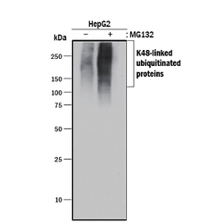
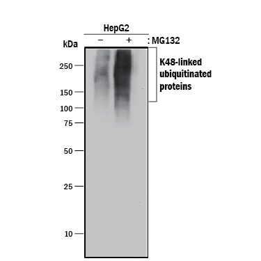
- Detection of Human Ubiquitin by Western Blot. Western blot shows lysates of HepG2 human hepatocellular carcinoma cell line untreated (-) or treated (+) with MG132. PVDF membrane was probed with 0.5 µg/mL of Rabbit Anti-Ubiquitin K48 Linkage Monoclonal Antibody (Catalog # A-101) followed by HRP-conjugated Anti-Rabbit IgG Secondary Antibody (Catalog # HAF008). A specific band was detected for Ubiquitin at approximately 75-250 kDa (as indicated). This experiment was conducted under reducing conditions and using Immunoblot Buffer Group 1.
- Submitted by
- R&D Systems (provider)
- Main image

- Experimental details
- Western Blot 25 ng of each linkage of recombinant diubiquitin was run on a 10-20% SDS-PAGE gel prior to blotting on PVDF membrane. Western blots were developed using anti-K48 mAb (A-101, upper panel) or anti-ubiquitin mAb (MAB701, lower panel) primaries at 0.5 µg/ml. The appropriate HRP-labeled anti-rabbit or anti-mouse (R&D Systems HAF008 or HAF007) secondary antibodies were used at a 1:2000 dilution. A single band of appropriate size was detected only in the K48-linked diubiquitin lane using A-101.
