Explore
 Validate
 Learn
Learn Western blot
 Immunocytochemistry
Antibody data
- Antibody Data
- Antigen structure
- References [6]
- Comments [0]
- Validations
- Western blot [2]
- Immunohistochemistry [1]
Submit
Validation data
Reference
Comment
Report error
- Product number
- NB300-129 - Provider product page

- Provider
- Novus Biologicals
- Proper citation
- Novus Cat#NB300-129, RRID:AB_2180545
- Product name
- Rabbit Polyclonal Ubiquitin Antibody
- Antibody type
- Polyclonal
- Description
- Unpurified. This antibody recognizes ubiquitinated inclusion bodies and both the mono- and polyubiquitin forms. This recognizes aggresomes.
- Reactivity
- Human, Mouse, Rat
- Host
- Rabbit
- Isotype
- IgG
- Vial size
- 0.05 ml
- Storage
- Store at 4C short term. Aliquot and store at -20C long term. Avoid freeze-thaw cycles.
Submitted references Profiling and identification of new proteins involved in brain ischemia using MALDI-imaging-mass-spectrometry.
Analysis of β-N-methylamino-L-alanine (L-BMAA) neurotoxicity in rat cerebellum.
CGG-repeat length threshold for FMR1 RNA pathogenesis in a cellular model for FXTAS.
Regulation of phosphoglucose isomerase/autocrine motility factor activities by the poly(ADP-ribose) polymerase family-14.
Protein composition of the intranuclear inclusions of FXTAS.
Induction of inclusion formation and disruption of lamin A/C structure by premutation CGG-repeat RNA in human cultured neural cells.
Llombart V, Trejo SA, Bronsoms S, Morancho A, Feifei M, Faura J, García-Berrocoso T, Simats A, Rosell A, Canals F, Hernández-Guillamón M, Montaner J
Journal of proteomics 2017 Jan 30;152:243-253
Journal of proteomics 2017 Jan 30;152:243-253
Analysis of β-N-methylamino-L-alanine (L-BMAA) neurotoxicity in rat cerebellum.
Muñoz-Sáez E, de Munck García E, Arahuetes Portero RM, Martínez A, Solas Alados MT, Miguel BG
Neurotoxicology 2015 May;48:192-205
Neurotoxicology 2015 May;48:192-205
CGG-repeat length threshold for FMR1 RNA pathogenesis in a cellular model for FXTAS.
Hoem G, Raske CR, Garcia-Arocena D, Tassone F, Sanchez E, Ludwig AL, Iwahashi CK, Kumar M, Yang JE, Hagerman PJ
Human molecular genetics 2011 Jun 1;20(11):2161-70
Human molecular genetics 2011 Jun 1;20(11):2161-70
Regulation of phosphoglucose isomerase/autocrine motility factor activities by the poly(ADP-ribose) polymerase family-14.
Yanagawa T, Funasaka T, Tsutsumi S, Hu H, Watanabe H, Raz A
Cancer research 2007 Sep 15;67(18):8682-9
Cancer research 2007 Sep 15;67(18):8682-9
Protein composition of the intranuclear inclusions of FXTAS.
Iwahashi CK, Yasui DH, An HJ, Greco CM, Tassone F, Nannen K, Babineau B, Lebrilla CB, Hagerman RJ, Hagerman PJ
Brain : a journal of neurology 2006 Jan;129(Pt 1):256-71
Brain : a journal of neurology 2006 Jan;129(Pt 1):256-71
Induction of inclusion formation and disruption of lamin A/C structure by premutation CGG-repeat RNA in human cultured neural cells.
Arocena DG, Iwahashi CK, Won N, Beilina A, Ludwig AL, Tassone F, Schwartz PH, Hagerman PJ
Human molecular genetics 2005 Dec 1;14(23):3661-71
Human molecular genetics 2005 Dec 1;14(23):3661-71
No comments: Submit comment
Supportive validation
- Submitted by
- Novus Biologicals (provider)
- Main image
- Experimental details
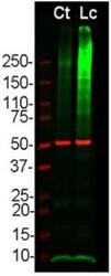
- Western Blot: Ubiquitin Antibody [NB300-129] - Analysis of Hek 293 cell lysates using Rabbit pAb to ubiquitin (1:5000 dilution, green) and Mouse mAb to B-Tubulin (1:10000 dilution, red) used as a loading control. Cells were maintained in normal medium (Control-Ct) or treated with proteasome inhibitor lactacystin (Lc) at 10 um for 16 h. Lysed cells were electrophoresed on 4-20% SDS-PAGE, and blotted to PVDF membrane. Note the smeary-patterned reactivities detected above the 200kDa standard that presumably represent accumulation of ubiquitinated proteins in proteasome inhibitor-Lc treated cells. Prominent band at 8kDa corresponds to monoubiquitin
- Submitted by
- Novus Biologicals (provider)
- Main image
- Experimental details
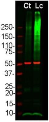
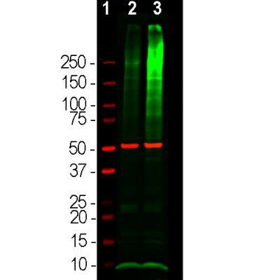
- Western Blot: Ubiquitin Antibody [NB300-129] - Analysis of HEK293 cell lysates using rabbit Ubiquitin antibody, dilution 1:5000 (Green), and mouse beta-Tubulin antibody, dilution 1:10000 (Red) used as a loading control. [1] protein standard (Red), [2] cells maintained in normal medium, [3] cells treated with proteasome inhibitor lactacystin (Lc) at 10uM for 16 hours. Lysed cells were lysed and the lysate subjected to electrophoresis on a 4-20% SDS-PAGE gel, then electrophoretically transferred to PVDF membranes. The smear detected above the 200kDa standard represents accumulations of ubiquitinated proteins in the Lc treated cells. The prominent band at ~8kDa corresponds to monoubiquitin.
Supportive validation
- Submitted by
- Novus Biologicals (provider)
- Main image
- Experimental details
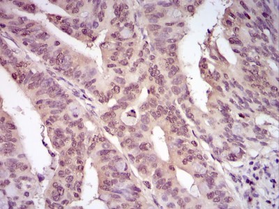
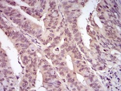
- Immunohistochemistry-Paraffin: Ubiquitin Antibody [NB300-129] - IHC staining of Ubiquitin in human rectal cancer using DAB with hematoxylin counterstain.