 Explore
 Validate
 Learn
LearnPA5-25584
antibody from Invitrogen Antibodies
Targeting: MARCHF5
FLJ20445, MARCH-V, MARCH5, MITOL, RNF153
 Western blot
 Immunohistochemistry
Antibody data
- Antibody Data
- Antigen structure
- References [1]
- Comments [0]
- Validations
- Immunohistochemistry [1]
- Flow cytometry [1]
- Other assay [1]
Submit
Validation data
Reference
Comment
Report error
- Product number
- PA5-25584 - Provider product page

- Provider
- Invitrogen Antibodies
- Product name
- MARCH5 Polyclonal Antibody
- Antibody type
- Polyclonal
- Antigen
- Synthetic peptide
- Description
- This antibody is predicted to react with bovine, chicken and Xenopus based on sequence homology.
- Reactivity
- Human, Mouse
- Host
- Rabbit
- Isotype
- IgG
- Vial size
- 400 μL
- Concentration
- 0.50 mg/mL
- Storage
- Store at 4°C short term. For long term storage, store at -20°C, avoiding freeze/thaw cycles.
Submitted references Parkin is a disease modifier in the mutant SOD1 mouse model of ALS.
Palomo GM, Granatiero V, Kawamata H, Konrad C, Kim M, Arreguin AJ, Zhao D, Milner TA, Manfredi G
EMBO molecular medicine 2018 Oct;10(10)
EMBO molecular medicine 2018 Oct;10(10)
No comments: Submit comment
Supportive validation
- Submitted by
- Invitrogen Antibodies (provider)
- Main image
- Experimental details
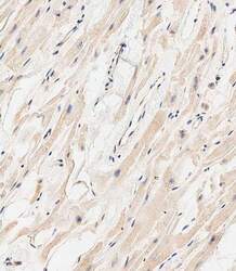
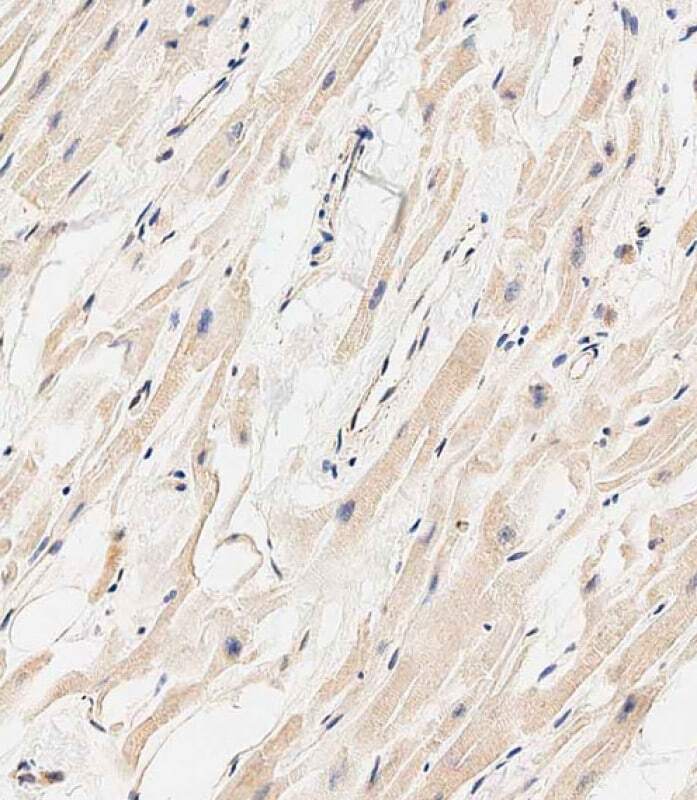
- Immunohistochemistry analysis of MARCH5 in paraffin-embedded Human heart tissue. Samples were incubated with MARCH5 polyclonal antibody (Product # PA5-25584) using a dilution of 1:500 for 1 hour at room temperature followed by an undiluted biotinylated CRF Anti-Polyvalent HRP Polymer antibody.
Supportive validation
- Submitted by
- Invitrogen Antibodies (provider)
- Main image
- Experimental details
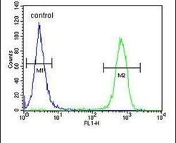
- Flow cytometry analysis of MCF-7 cells using a MARCH5 polyclonal antibody (Product # PA5-25584) (right) compared to a negative control cell (left) at a dilution of 1:10-50, followed by a FITC-conjugated goat anti-rabbit antibody

Supportive validation
- Submitted by
- Invitrogen Antibodies (provider)
- Main image
- Experimental details
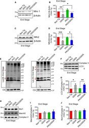
- Figure 8 Parkin knockout improves the decline of mitochondrial dynamics proteins in SOD 1-G93A spinal cord, but mitochondrial protein ubiquitination profiles and March5 and Mul1 levels are unaffected A, B Western blots (A) and quantification (B) of Miro1 in spinal cord homogenates at disease end stage. beta-actin is used as loading reference. Results are expressed as mean +- SEM and as percent of Non Tg; n = 8 (four males and four females) mice per group; * P = 0.011 by paired Student's t -test (for comparison G93A vs. PKO/G93A) and ** P = 0.003 by paired Friedman's test with Dunn's correction (for Non Tg vs. G93A). Parkin knockout increases the levels of Miro1 in SOD1-G93A mice at disease end stage. C, D Western blots (C) and quantification (D) of Mfn2 in spinal cord homogenates at disease end stage. Protein levels are normalized by beta-actin. Results are expressed as mean +- SEM and as percent of Non Tg; n = 8 (four males and four females) mice per group. No statistically significant differences were found between G93A and PKO/G93A ( P = 0.078 by paired Wilcoxon's test); *** P = 0.0007 by paired Friedman's test with Dunn's correction (for Non Tg vs. G93A) and * P = 0.037 by paired Friedman's test with Dunn's correction (for PKO vs. PKO/G93A). Parkin knockout increases the levels of Mfn2 in SOD1-G93A mice at disease end stage. E Western blots of spinal cord mitochondria at disease end stage probed for lysine 48 (left panel) and lysine 63 (right panel) ubiquitin chains. C
