Explore
 Validate
 Learn
Learn Western blot
 Immunohistochemistry
 Other assay
Antibody data
- Antibody Data
- Antigen structure
- References [2]
- Comments [0]
- Validations
- Other assay [3]
Submit
Validation data
Reference
Comment
Report error
- Product number
- PA5-28522 - Provider product page

- Provider
- Invitrogen Antibodies
- Product name
- SPOP Polyclonal Antibody
- Antibody type
- Polyclonal
- Antigen
- Recombinant full-length protein
- Description
- Recommended positive controls: HepG2, HepG2 nuclear extract, Mouse brain, SPOP-transfected 293T. Predicted reactivity: Mouse (100%), Rat (100%), Zebrafish (99%), Xenopus laevis (100%), Rhesus Monkey (100%), Bovine (100%). Store product as a concentrated solution. Centrifuge briefly prior to opening the vial.
- Reactivity
- Human, Mouse
- Host
- Rabbit
- Isotype
- IgG
- Vial size
- 100 μL
- Concentration
- 0.1 mg/mL
- Storage
- Store at 4°C short term. For long term storage, store at -20°C, avoiding freeze/thaw cycles.
Submitted references Impaired Proteolysis of Noncanonical RAS Proteins Drives Clonal Hematopoietic Transformation.
T-box3 is a ciliary protein and regulates stability of the Gli3 transcription factor to control digit number.
Chen S, Vedula RS, Cuevas-Navarro A, Lu B, Hogg SJ, Wang E, Benbarche S, Knorr K, Kim WJ, Stanley RF, Cho H, Erickson C, Singer M, Cui D, Tittley S, Durham BH, Pavletich TS, Fiala E, Walsh MF, Inoue D, Monette S, Taylor J, Rosen N, McCormick F, Lindsley RC, Castel P, Abdel-Wahab O
Cancer discovery 2022 Oct 5;12(10):2434-2453
Cancer discovery 2022 Oct 5;12(10):2434-2453
T-box3 is a ciliary protein and regulates stability of the Gli3 transcription factor to control digit number.
Emechebe U, Kumar P P, Rozenberg JM, Moore B, Firment A, Mirshahi T, Moon AM
eLife 2016 Apr 5;5
eLife 2016 Apr 5;5
No comments: Submit comment
Supportive validation
- Submitted by
- Invitrogen Antibodies (provider)
- Main image
- Experimental details
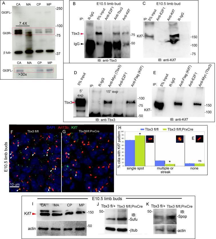
- Figure 4. Loss of Tbx3 results in Gli3 protein instability and aberrant localization of Kif7 in limb bud cilia. ( A ) Representative immunoblot (N=3) blot of E10.75 forelimb bud lysates prepared from microdissected Tbx3 fl/+ control anterior limb (CA), Tbx3;PrxCre mutant anterior limb (MA), control posterior (CP), and mutant posterior (MP) probed for Gli3 and betatubulin loading control. Note decreased level of Gli3FL and Gli3R, and multiple bands of lower molecular weight than Gli3R in MA sample. Densitometry of Gli3R bands in red box in N revealed that in this representative experiment, the level of Gli3R was 7.4 fold decreased in mutant anterior relative to control anterior. ( A' ) Longer exposure of top of blot shown in panel A to examine Gli3FL band. The control (CA) Gli3FL band is 31 fold more intense than mutant (MA, virtually undetectable). ( B ) Immunoblot of lysates from E10.5 forelimb buds immunoprecipitated (IB) with antibodies listed at top and immunoblotted (IB) for Tbx3. Lane 5 shows that immunoprecipitation with anti-Kif7 antibody co-IPs Tbx3. ( C ) As in panel B, but assayed for Kif7. ( D ) Co-IP assay of Myc-tagged Tbx3 and Flag-tagged GFP overexpressed in HEK293 cells. IP was performed with antibodies listed at top and immunoblotted for Tbx3. Myc-tagged Tbx3 co-IPs with Flag-tagged Kif7. Input lane was 5 s exposure (5"" exp) while other lanes were 15 s (15"" exp). ( E ) As in D , but in this case, blot probed for Kif7; confirms interaction of tagged, o
- Submitted by
- Invitrogen Antibodies (provider)
- Main image
- Experimental details
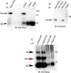
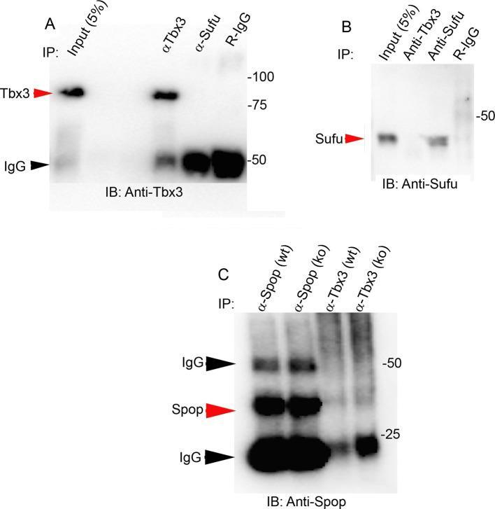
- Figure 7--figure supplement 3. Tbx3 does not co-IP with Sufu or Spop in mouse embryo lysates. ( A , B ) Immunoprecipitations/Immunoblot assaying for interaction between endogenous Tbx3 and Sufu in E10.5 mouse embryo lysates. Sufu did not co-IP Tbx3 ( A ) nor did Tbx3 co-IP Sufu ( B ). ( C ) Immunoprecipitations/Immunoblot assaying for interaction between Tbx3 and Spop in control and Tbx3 Deltafl/Deltafl E10.5 mouse embryo lysates. No interaction was detected. Note increased Spop in mutant, consistent with previous result in Figure 4J and data in Figure 8--figure supplement 1 . DOI: http://dx.doi.org/
- Submitted by
- Invitrogen Antibodies (provider)
- Main image

- Experimental details
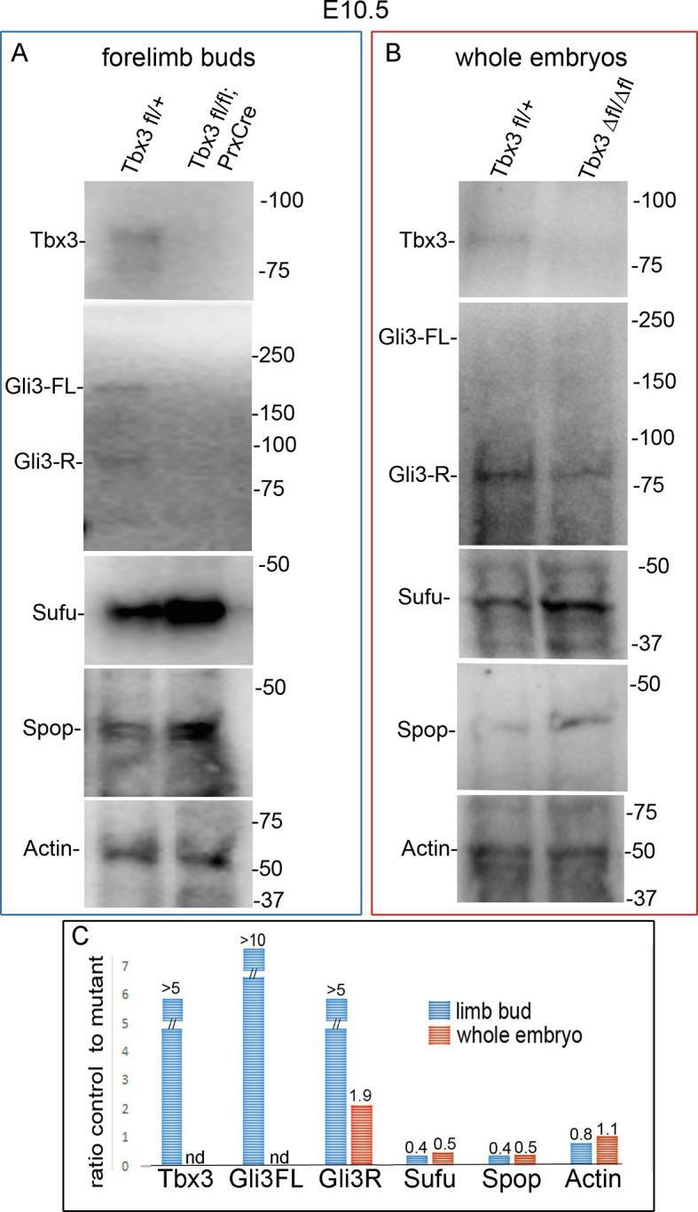
- Figure 8--figure supplement 1. Altered protein levels observed in mutant limb buds are also apparent in whole embryos. ( A , B ) Immunoblots on protein lysates prepared from E10.5 forelimb buds ( A ) and whole embryos ( B ). Actin is loading control. ( C ) Quantitation of protein levels in A and B comparing amount detected in control to mutant. Note increased levels of Sufu and Spop in mutant limbs and embryos that results in ratios of control to mutant