 Explore
 Validate
 Learn
Learn Western blot
 Immunocytochemistry
Antibody data
- Antibody Data
- Antigen structure
- References [0]
- Comments [0]
- Validations
- Immunocytochemistry [2]
- Flow cytometry [3]
Submit
Validation data
Reference
Comment
Report error
- Product number
- MA5-24251 - Provider product page

- Provider
- Invitrogen Antibodies
- Product name
- Galectin 10 Monoclonal Antibody (561603)
- Antibody type
- Monoclonal
- Antigen
- Recombinant full-length protein
- Description
- In direct ELISAs, no cross-reactivity with recombinant human Galectin-1, -2, -3, -4, -7, -8, or -9/Ecalectin is observed. Reconstitute in sterile PBS to a final concentration of 0.5 mg/mL.
- Reactivity
- Human
- Host
- Mouse
- Isotype
- IgG
- Antibody clone number
- 561603
- Vial size
- 100 μg
- Concentration
- 0.5 mg/mL
- Storage
- -20°C, Avoid Freeze/Thaw Cycles
No comments: Submit comment
Supportive validation
- Submitted by
- Invitrogen Antibodies (provider)
- Main image

- Experimental details
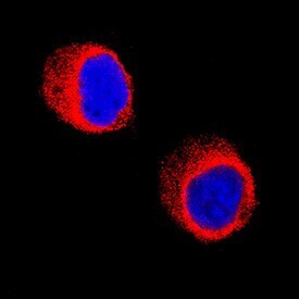
- Immunocytochemistry analysis of Galectin 10 in immersion fixed HL‚60 human acute promyelocytic leukemia cell line. Samples were incubated in Galectin 10 monoclonal antibody (Product # MA5-24251) using a dilution of 8 µg/mL for 3 hours at room temperature followed by NorthernLights™ 557-conjugated Anti-Mouse IgG Secondary Antibody (red) and counterstained with DAPI (blue). Specific staining was localized to cytoplasm.

- Submitted by
- Invitrogen Antibodies (provider)
- Main image

- Experimental details
- Immunocytochemistry analysis of Galectin 10 in immersion fixed HL‚60 human acute promyelocytic leukemia cell line. Samples were incubated in Galectin 10 monoclonal antibody (Product # MA5-24251) using a dilution of 8 µg/mL for 3 hours at room temperature followed by NorthernLights™ 557-conjugated Anti-Mouse IgG Secondary Antibody (red) and counterstained with DAPI (blue). Specific staining was localized to cytoplasm.
Supportive validation
- Submitted by
- Invitrogen Antibodies (provider)
- Main image
- Experimental details
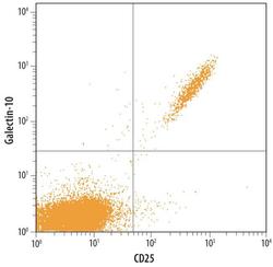
- Flow cytometric analysis of human peripheral blood lymphocytes were stained with Human Galectin-10 Monoclonal Antibody (Product # MA5-24251) followed by FITC-conjugated Anti-Mouse IgG Secondary Antibody and Human IL-2Rα APC-conjugated Monoclonal Antibody. Quadrant markers were set based on control antibody staining.

- Submitted by
- Invitrogen Antibodies (provider)
- Main image

- Experimental details
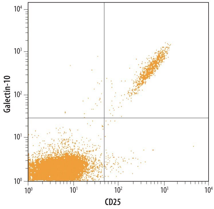
- Flow cytometry of Galectin 10 in Human peripheral blood lymphocytes. Samples were incubated in Galectin 10 monoclonal antibody (Product # MA5-24251) followed by Fluorescein-conjugated Anti-Mouse IgG Secondary Antibody and Human IL‚2 Rα APC-conjugated Monoclonal Antibody. Quadrant markers were set based on control antibody staining.
- Submitted by
- Invitrogen Antibodies (provider)
- Main image

- Experimental details
- Flow cytometry of Galectin 10 in Human peripheral blood lymphocytes. Samples were incubated in Galectin 10 monoclonal antibody (Product # MA5-24251) followed by Fluorescein-conjugated Anti-Mouse IgG Secondary Antibody and Human IL‚2 Rα APC-conjugated Monoclonal Antibody. Quadrant markers were set based on control antibody staining.
