Explore
 Validate
 Learn
Learn Western blot
Antibody data
- Antibody Data
- Antigen structure
- References [0]
- Comments [0]
- Validations
- Western blot [1]
- Immunohistochemistry [5]
Submit
Validation data
Reference
Comment
Report error
- Product number
- PA5-105874 - Provider product page

- Provider
- Invitrogen Antibodies
- Product name
- Phospho-DYRK1A/DYRK1B (Tyr321, Tyr273) Polyclonal Antibody
- Antibody type
- Polyclonal
- Antigen
- Synthetic peptide
- Description
- Antibody detects endogenous levels of DYRK1A/B only when phosphorylated at Tyr321/273.
- Reactivity
- Human, Mouse, Rat
- Host
- Rabbit
- Isotype
- IgG
- Vial size
- 100 μL
- Concentration
- 1 mg/mL
- Storage
- -20°C
No comments: Submit comment
Supportive validation
- Submitted by
- Invitrogen Antibodies (provider)
- Main image
- Experimental details
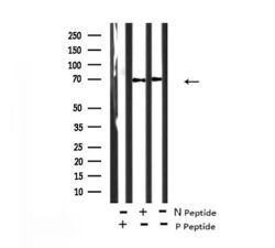
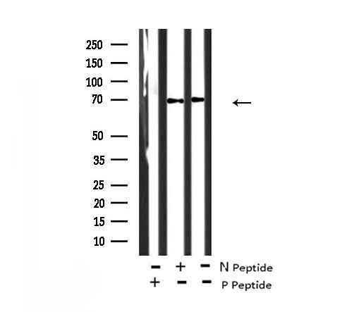
- Western blot analysis of Phospho-DYRK1A/DYRK1B (Tyr321, Tyr273) in EGF treated K562 cell lysate (absence (-) or presence (+) of non-phospho and phospho peptide). Samples were incubated with Phospho-DYRK1A/DYRK1B (Tyr321, Tyr273) polyclonal antibody (Product # PA5-105874).
Supportive validation
- Submitted by
- Invitrogen Antibodies (provider)
- Main image
- Experimental details
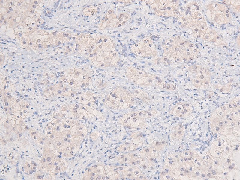
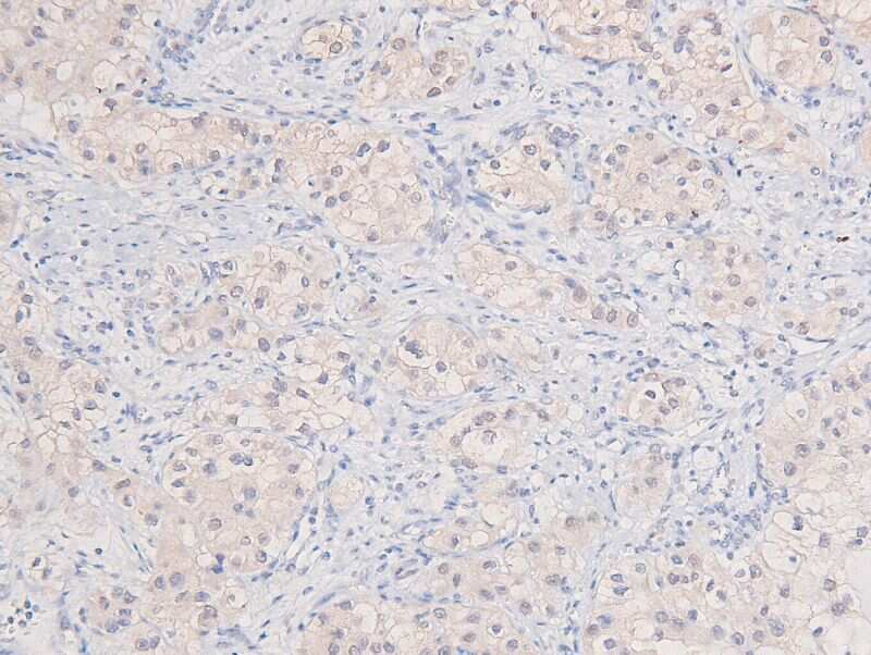
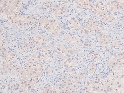
- Immunohistochemistry analysis of paraffin-embedded Phospho-DYRK1A/DYRK1B (Tyr321, Tyr273) in human liver cancer tissue sections. Antigen retrieval was performed using citrate buffer. Samples were blocked with blocking buffer (1.5 hr, 22°C), incubated with Phospho-DYRK1A/DYRK1B (Tyr321, Tyr273) polyclonal antibody (Product # PA5-105874) using a dilution of 1:200 (1.5 hr, 22°C), followed by HRP conjugated goat anti-rabbit.
- Submitted by
- Invitrogen Antibodies (provider)
- Main image
- Experimental details
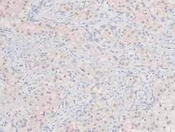
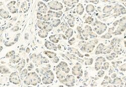
- Immunohistochemistry analysis of Phospho-DYRK1A/DYRK1B (Tyr321, Tyr273) in human normal tissues adjacent to pancreatic cancer. The sample was formaldehyde fixed and a heat mediated antigen retrieval step in citrate buffer was performed. Samples were incubated with Phospho-DYRK1A/DYRK1B (Tyr321, Tyr273) polyclonal antibody (Product # PA5-105874) using a dilution of 1:100 (4°C overnight) followed by HRP conjugated anti-Rabbit secondary antibody.

- Submitted by
- Invitrogen Antibodies (provider)
- Main image
- Experimental details
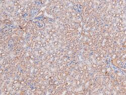
- Immunohistochemistry analysis of paraffin-embedded Phospho-DYRK1A/DYRK1B (Tyr321, Tyr273) in mouse kidney tissue sections. Antigen retrieval was performed using citrate buffer. Samples were blocked with blocking buffer (1.5 hr, 22°C), incubated with Phospho-DYRK1A/DYRK1B (Tyr321, Tyr273) polyclonal antibody (Product # PA5-105874) using a dilution of 1:200 (1.5 hr, 22°C), followed by HRP conjugated goat anti-rabbit.

- Submitted by
- Invitrogen Antibodies (provider)
- Main image
- Experimental details
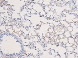
- Immunohistochemistry analysis of paraffin-embedded Phospho-DYRK1A/DYRK1B (Tyr321, Tyr273) in rat lung tissue sections. Antigen retrieval was performed using citrate buffer. Samples were blocked with blocking buffer (1.5 hr, 22°C), incubated with Phospho-DYRK1A/DYRK1B (Tyr321, Tyr273) polyclonal antibody (Product # PA5-105874) using a dilution of 1:200 (1.5 hr, 22°C), followed by HRP conjugated goat anti-rabbit.

- Submitted by
- Invitrogen Antibodies (provider)
- Main image
- Experimental details
- Immunohistochemistry analysis of paraffin-embedded Phospho-DYRK1A/DYRK1B (Tyr321, Tyr273) in human liver cancer tissue sections. Antigen retrieval was performed using citrate buffer. Samples were blocked with blocking buffer (1.5 hr, 22°C), incubated with Phospho-DYRK1A/DYRK1B (Tyr321, Tyr273) polyclonal antibody (Product # PA5-105874) using a dilution of 1:200 (1.5 hr, 22°C), followed by HRP conjugated goat anti-rabbit.