Explore
 Validate
 Learn
Learn Western blot
 ELISA
ELISAAntibody data
- Antibody Data
- Antigen structure
- References [3]
- Comments [0]
- Validations
- Western blot [1]
- Immunocytochemistry [2]
- Other assay [2]
Submit
Validation data
Reference
Comment
Report error
- Product number
- MA3-033 - Provider product page

- Provider
- Invitrogen Antibodies
- Product name
- CYP3A5 Monoclonal Antibody (F18 P3 B6)
- Antibody type
- Monoclonal
- Antigen
- Synthetic peptide
- Description
- MA3-033 detects Cytochrome P450 3A5 from HEK293 and HepG2 human cell lines, and from bovine liver and pancreas samples. MA3-033 has been successfully used in Western blot applications. By Western blot, this antibody detects a ~54 kDa band representing Cytochrome P450 3A5. The MA3-033 immunogen is a synthetic peptide corresponding to residues E S R D G T L S G E, conjugated to ovalbumin.
- Reactivity
- Human, Bovine
- Host
- Mouse
- Isotype
- IgG
- Antibody clone number
- F18 P3 B6
- Vial size
- 100 μL
- Concentration
- Conc. Not Determined
- Storage
- -20°C, Avoid Freeze/Thaw Cycles
Submitted references Antisense oligonucleotide development for the selective modulation of CYP3A5 in renal disease.
Role of CYP3A5 in Modulating Androgen Receptor Signaling and Its Relevance to African American Men with Prostate Cancer.
Cytochrome p450 profile of colorectal cancer: identification of markers of prognosis.
Lidberg KA, Annalora AJ, Jozic M, Elson DJ, Wang L, Bammler TK, Ramm S, Monteiro MB, Himmelfarb J, Marcus CB, Iversen PL, Kelly EJ
Scientific reports 2021 Feb 25;11(1):4722
Scientific reports 2021 Feb 25;11(1):4722
Role of CYP3A5 in Modulating Androgen Receptor Signaling and Its Relevance to African American Men with Prostate Cancer.
Gorjala P, Kittles RA, Goodman OB Jr, Mitra R
Cancers 2020 Apr 17;12(4)
Cancers 2020 Apr 17;12(4)
Cytochrome p450 profile of colorectal cancer: identification of markers of prognosis.
Kumarakulasingham M, Rooney PH, Dundas SR, Telfer C, Melvin WT, Curran S, Murray GI
Clinical cancer research : an official journal of the American Association for Cancer Research 2005 May 15;11(10):3758-65
Clinical cancer research : an official journal of the American Association for Cancer Research 2005 May 15;11(10):3758-65
No comments: Submit comment
Supportive validation
- Submitted by
- Invitrogen Antibodies (provider)
- Main image
- Experimental details
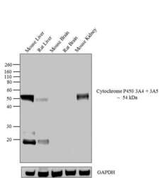
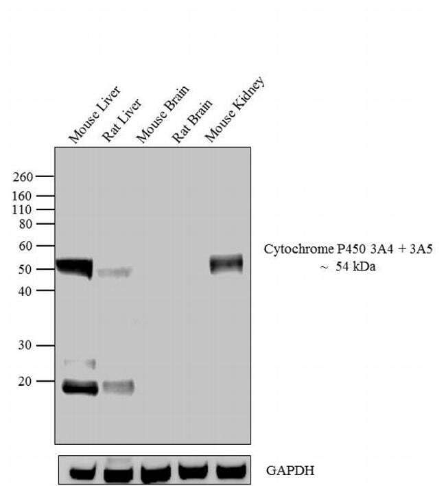
- Western blot analysis was performed on whole cell extracts (30 µg lysate) of Mouse Liver (Lane 1), Rat Liver (Lane 2), Mouse Brain (Lane 3), Rat Brain (Lane 4) and Mouse Kidney (Lane 5). The blots were probed with Anti-Cytochrome P450 3A5 Mouse Monoclonal Antibody (Product # MA3-033, 1:100-1:1000 dilution) and detected by chemiluminescence using Goat anti-Mouse IgG (H+L) Secondary Antibody, HRP conjugate (Product # 62-6520, 1:4000 dilution). A 54 kDa band corresponding to Cytochrome P450 3A4 + 3A5 was observed across cell lines tested except in Mouse Brain and Rat Brain. An extra band was observed at approximately ~ 20 kDa in Mouse Liver and Rat Liver. Known quantity of protein samples were electrophoresed using Novex® NuPAGE® 10 % Bis-Tris gel (Product # NP0301BOX), XCell SureLock™ Electrophoresis System (Product # EI0002) and Novex® Sharp Pre-Stained Protein Standard (Product # LC5800). Resolved proteins were then transferred onto a nitrocellulose membrane with iBlot® 2 Dry Blotting System (Product # IB21001). The membrane was probed with the relevant primary and secondary Antibody following blocking with 5 % skimmed milk. Chemiluminescent detection was performed using Pierce™ ECL Western Blotting Substrate (Product # 32106).
Supportive validation
- Submitted by
- Invitrogen Antibodies (provider)
- Main image

- Experimental details
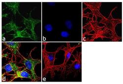
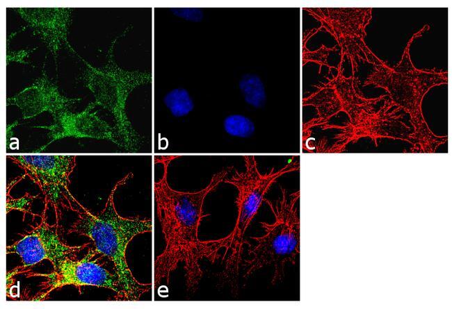
- Immunofluorescence analysis of Cytochrome P450-3A5 was done on 70% confluent log phase HepG2 cells. The cells were fixed with 4% paraformaldehyde for 10 minutes, permeabilized with 0.1% Triton™ X-100 for 10 minutes, and blocked with 1% BSA for 1 hour at room temperature. The cells were labeled with Cytochrome P450 3A5 (F18 P3 B6) Mouse Monoclonal Antibody (Product # MA3-033) at 1:250 dilution in 0.1% BSA and incubated for 3 hours at room temperature and then labeled with Goat anti-Mouse IgG (H+L) Superclonal™ Secondary Antibody, Alexa Fluor® 488 conjugate (Product # A28175) at a dilution of 1:2000 for 45 minutes at room temperature (Panel a: green). Nuclei (Panel b: blue) were stained with SlowFade® Gold Antifade Mountant with DAPI (Product # S36938). F-actin (Panel c: red) was stained with Rhodamine Phalloidin (Product # R415, 1:300). Panel d is a merged image showing cytoplasmic localization. Panel e is a no primary antibody control. The images were captured at 60X magnification.

- Submitted by
- Invitrogen Antibodies (provider)
- Main image
- Experimental details
- Immunofluorescence analysis of Cytochrome P450-3A5 was done on 70% confluent log phase HepG2 cells. The cells were fixed with 4% paraformaldehyde for 10 minutes, permeabilized with 0.1% Triton™ X-100 for 10 minutes, and blocked with 1% BSA for 1 hour at room temperature. The cells were labeled with Cytochrome P450 3A5 (F18 P3 B6) Mouse Monoclonal Antibody (Product # MA3-033) at 1:250 dilution in 0.1% BSA and incubated for 3 hours at room temperature and then labeled with Goat anti-Mouse IgG (H+L) Superclonal™ Secondary Antibody, Alexa Fluor® 488 conjugate (Product # A28175) at a dilution of 1:2000 for 45 minutes at room temperature (Panel a: green). Nuclei (Panel b: blue) were stained with SlowFade® Gold Antifade Mountant with DAPI (Product # S36938). F-actin (Panel c: red) was stained with Rhodamine Phalloidin (Product # R415, 1:300). Panel d is a merged image showing cytoplasmic localization. Panel e is a no primary antibody control. The images were captured at 60X magnification.
Supportive validation
- Submitted by
- Invitrogen Antibodies (provider)
- Main image
- Experimental details
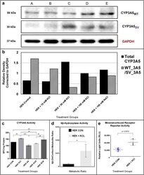
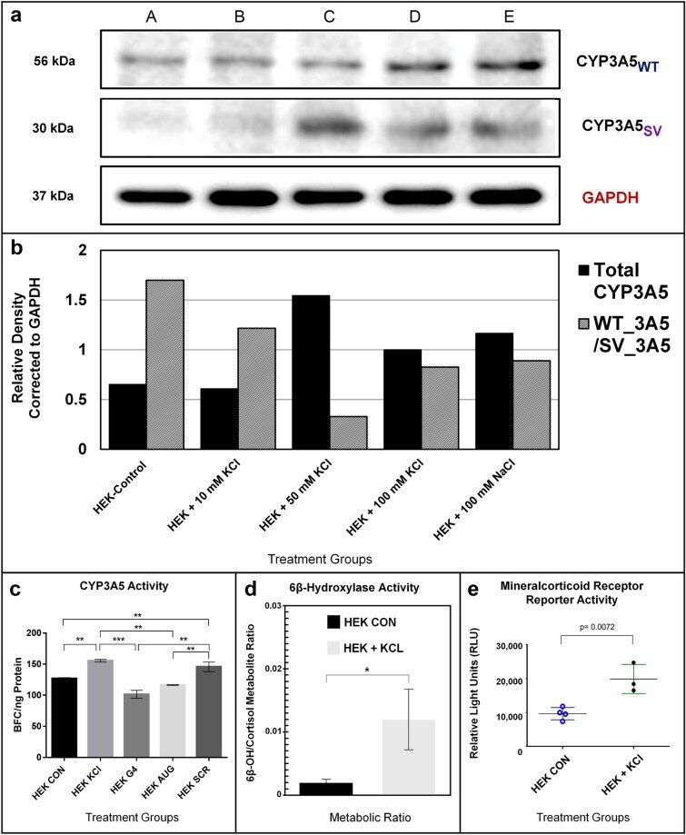
- Figure 4 Salt-Induced CYP3A5*3 Protein Expression, Enzyme Activity and Mineralocorticoid Receptor Signaling in HEK293 Cells. ( a ) Confluent HEK293 cells were exposed for 48 h to (A) control media or media supplemented with (B) 10 mM KCl, (C) 50 mM KCl, (D) 100 mM KCl, or (E) 100 mM NaCl. Two protein bands attributed to CYP3A5 were detected via western blot, including the full-length reference protein ( CYP3A5 WT ) at 56 kilodaltons (kDa) and a truncated 30 kDa splice variant form ( CYP3A5 sv ). The housekeeping gene GAPDH , detected at 37 kDa, was used to normalize sample loading. ( b ) Total CYP3A5 protein and the ratio of CYP3A5 WT to CYP3A5 SV protein normalized to GAPDH expression was determined using densiometric analysis in ImageJ. ( c ) CYP3A enzyme activity was monitored in HEK293 cells under normal conditions and with elevated KCl (12 mM; 48 h.) and PMOs (1 µM; 48 h. ), as measured fluorometrically by BFC metabolism (see "" Methods ""). KCl supplementation significantly induced enzyme activity ( p < 0.01; t-test) in HEK293 cells (KCl) compared to control cells (CON). The G4 disrupting PMO (G4) and the CYP3A5 start-site inhibitor (AUG) PMO both significantly prevented KCl induction of BFC metabolism ( p < 0.01 compared to KCl treated control) and were not different from control cells. The scrambled (SCR) PMO control did not inhibit KCl induction of CYP3A activity (ANOVA p value < 0.01. Post test p < 0.01 = **, and p < 0.005 = ***). ( d ) LC MS/MS analysis of HE
- Submitted by
- Invitrogen Antibodies (provider)
- Main image

- Experimental details
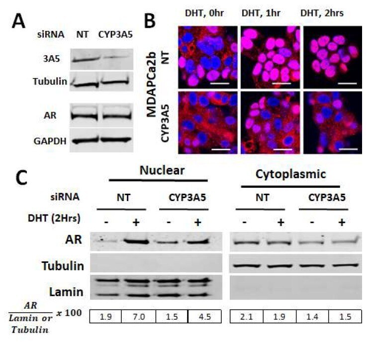
- Figure 1 CYP3A5 siRNA downregulates AR (androgen receptor) nuclear translocation: ( A - C ) MDAPCa2b cells were transfected with CYP3A5 and non target (NT) siRNA. After 72 hours, the cells were given 10nM DHT treatment (0, 1, and 2 hours). ( A ) Western blot was performed to test CYP3A5 siRNA silencing efficiency at protein level using cytoskeletal fraction. Total protein was used to monitor changes in total AR protein expression. ( B ) For microscopy, the cells were labelled with AR primary antibody and Cy5 secondary (red) and nucleus was labeled with DAPI. The scale bar represents 50 um. ( C ) After cell fractionation, western blotting was performed using cytoplasmic and nuclear fractions and probed for AR, Tubulin, and Lamin.