 Explore
 Validate
 Learn
Learn Western blot
Antibody data
- Antibody Data
- Antigen structure
- References [0]
- Comments [0]
- Validations
- Western blot [1]
- Immunocytochemistry [1]
Submit
Validation data
Reference
Comment
Report error
- Product number
- MA3-032 - Provider product page

- Provider
- Invitrogen Antibodies
- Product name
- CYP3A4/CYP3A5 Monoclonal Antibody (F24 P2 B10)
- Antibody type
- Monoclonal
- Antigen
- Synthetic peptide
- Description
- MA3-032 detects Cytochrome P450 3A4/3A5 from human cell line HepG2, and from bovine liver and pancreas samples. MA3-032 has been successfully used in Western blot applications. By Western blot, this antibody detects a ~54 kDa band representing Cytochrome P450 3A4/3A5. The MA3-032 immunogen is a synthetic peptide corresponding to residues E S R D G T V S G A, conjugated to ovalbumin.
- Reactivity
- Human, Bovine
- Host
- Mouse
- Isotype
- IgG
- Antibody clone number
- F24 P2 B10
- Vial size
- 100 µL
- Concentration
- Conc. Not Determined
- Storage
- -20° C, Avoid Freeze/Thaw Cycles
No comments: Submit comment
Supportive validation
- Submitted by
- Invitrogen Antibodies (provider)
- Main image

- Experimental details
- Western blot analysis was performed on whole cell extracts (30 µg lysate) of Mouse Liver (Lane 1), Rat Liver (Lane 2), Mouse Brain (Lane 3), Rat Brain (Lane 4) and Mouse Kidney (Lane 5). The blots were probed with Anti-Cytochrome P450 3A4 + 3A5 Mouse Monoclonal Antibody (Product # MA3-032, 1:100-1:1000 dilution) and detected by chemiluminescence using Goat anti-Mouse IgG (H+L) Secondary Antibody, HRP conjugate (Product # 62-6520, 1:4000 dilution). A 54 kDa band corresponding to Cytochrome P450 3A4 + 3A5 was observed across tissue lysates tested except in Mouse Brain and Rat Brain. An additional band was observed at ~ 23 kDa in Mouse Liver and Rat Liver. Known quantity of protein samples were electrophoresed using Novex® NuPAGE®10 % Bis-Tris gel (Product # NP0301BOX), XCell SureLock™ Electrophoresis System (Product # EI0002) and Novex® Sharp Pre-Stained Protein Standard (Product # LC5800). Resolved proteins were then transferred onto a nitrocellulose membrane with iBlot® 2 Dry Blotting System (Product # IB21001). The membrane was probed with the relevant primary and secondary Antibody following blocking with 5 % skimmed milk. Chemiluminescent detection was performed using Pierce™ ECL Western Blotting Substrate (Product # 32106).

Supportive validation
- Submitted by
- Invitrogen Antibodies (provider)
- Main image
- Experimental details
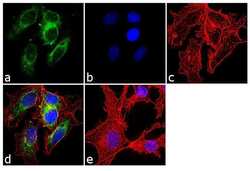
- Immunofluorescence analysis of Cytochrome P450-3A4-3A5 was done on 70% confluent log phase HepG2 cells. The cells were fixed with 4% paraformaldehyde for 10 minutes, permeabilized with 0.1% Triton™ X-100 for 10 minutes, and blocked with 1% BSA for 1 hour at room temperature. The cells were labeled with Cytochrome P450 3A4 + 3A5 (F24 P2 B10) Mouse Monoclonal Antibody (Product # MA3-032) at 1:250 dilution in 0.1% BSA and incubated for 3 hours at room temperature and then labeled with Goat anti-Mouse IgG (H+L) Superclonal™ Secondary Antibody, Alexa Fluor® 488 conjugate (Product # A28175) at a dilution of 1:2000 for 45 minutes at room temperature (Panel a: green). Nuclei (Panel b: blue) were stained with SlowFade® Gold Antifade Mountant with DAPI (Product # S36938). F-actin (Panel c: red) was stained with Rhodamine Phalloidin (Product # R415, 1:300). Panel d is a merged image showing cytoplasmic localization. Panel e is a no primary antibody control. The images were captured at 60X magnification.
