 Explore
 Validate
 Learn
Learn Western blot
Antibody data
- Antibody Data
- Antigen structure
- References [1]
- Comments [0]
- Validations
- Western blot [4]
- Immunocytochemistry [2]
Submit
Validation data
Reference
Comment
Report error
- Product number
- PA5-30048 - Provider product page

- Provider
- Invitrogen Antibodies
- Product name
- Golgin-97 Polyclonal Antibody
- Antibody type
- Polyclonal
- Antigen
- Recombinant full-length protein
- Description
- Recommended positive controls: A549, HeLa, HepG2, HCT116, mouse brain. Predicted reactivity: Mouse (90%), Rat (90%), Bovine (93%). Store product as a concentrated solution. Centrifuge briefly prior to opening the vial.
- Reactivity
- Human, Mouse, Hamster
- Host
- Rabbit
- Isotype
- IgG
- Vial size
- 100 μL
- Concentration
- 0.29 mg/mL
- Storage
- Store at 4°C short term. For long term storage, store at -20°C, avoiding freeze/thaw cycles.
Submitted references BECLIN1 Is Essential for Podocyte Secretory Pathways Mediating VEGF Secretion and Podocyte-Endothelial Crosstalk.
Bork T, Liang W, Kretz O, Lagies S, Yamahara K, Hernando-Erhard C, Helmstädter M, Schell C, Kammerer B, Huber TB
International journal of molecular sciences 2022 Mar 30;23(7)
International journal of molecular sciences 2022 Mar 30;23(7)
No comments: Submit comment
Supportive validation
- Submitted by
- Invitrogen Antibodies (provider)
- Main image
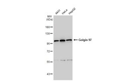
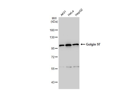
- Experimental details
- Western Blot using Golgin-97 Polyclonal Antibody (Product # PA5-30048). Various whole cell extracts (30 µg) were separated by 7.5% SDS-PAGE, and the membrane was blotted with Golgin-97 Polyclonal Antibody (Product # PA5-30048) diluted at 1:1,000. The HRP-conjugated anti-rabbit IgG antibody was used to detect the primary antibody.
- Submitted by
- Invitrogen Antibodies (provider)
- Main image

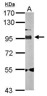
- Experimental details
- Western Blot using Golgin-97 Polyclonal Antibody (Product # PA5-30048). Sample (50 µg of whole cell lysate). Lane A: Mouse brain . 7.5% SDS PAGE. Golgin-97 Polyclonal Antibody (Product # PA5-30048) diluted at 1:1,000.
- Submitted by
- Invitrogen Antibodies (provider)
- Main image
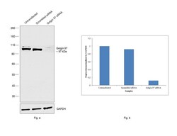
- Experimental details
- KD of Golgin-97 was achieved by transfecting T-47D with Golgin-97 specific siRNAs (Silencer® select Product # s5940, s2939). Western blot analysis (Fig. a) was performed using whole cell extracts from the Golgin-97 KD cells (Lane 3), non-specific scrambled siRNA transfected cells (Lane 2) and untransfected cells (Lane 1). The blot was probed with Golgin-97 Polyclonal Antibody (Product # PA5-30048, 1:500 dilution) and Goat anti-Rabbit IgG (H+L) Superclonal™ Secondary Antibody, HRP conjugate (Product # A27036, 1:4000 dilution). Densitometric analysis of this western blot is shown in histogram (Fig. b). Decrease in signal upon siRNA mediated knock down confirms that antibody is specific to Golgin-97..

- Submitted by
- Invitrogen Antibodies (provider)
- Main image
- Experimental details
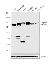
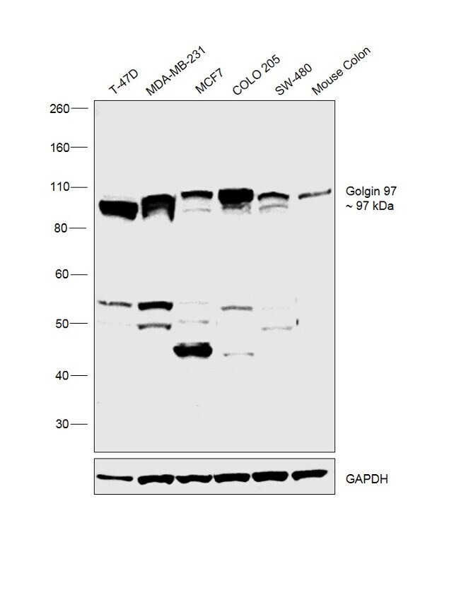
- Western blot was performed using Anti-Gologin-97 Polyclonal Antibody (Product # PA5-30048) and 97 kDa band corresponding to Golgin-97 was observed across cell lines and tissue tested. Whole cell extracts (30 µg lysate) of T-47D (Lane 1), MDA-MB-231 (Lane 2), MCF7 (Lane 3), COLO 205 (Lane 4), SW-480 (Lane 5) and tissue extract of Mouse Colon (Lane 6) were electrophoresed using Novex® NuPAGE® 4-12 % Bis-Tris gel (Product # NP0322BOX). Resolved proteins were then transferred onto a nitrocellulose membrane (Product # IB23001) by iBlot® 2 Dry Blotting System (Product # IB21001). The blot was probed with the primary antibody (1:500 dilution) and detected by Goat Anti-Rabbit IgG Secondary Antibody, HRP conjugate (Product # A27036, 1:4000 dilution) using the iBright FL 1000 (Product # A32752). Chemiluminescent detection was performed using Novex® ECL Chemiluminescent Substrate Reagent Kit (Product # WP20005). .
Supportive validation
- Submitted by
- Invitrogen Antibodies (provider)
- Main image
- Experimental details
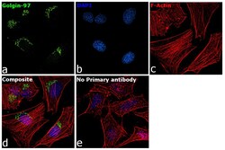
- Immunofluorescence analysis of Golgin-97 was performed using 70% confluent log phase HeLa cells. The cells were fixed with 4% paraformaldehyde for 10 minutes, permeabilized with 0.1% Triton™ X-100 for 15 minutes, and blocked with 2% BSA for 1 hour at room temperature. The cells were labeled with Golgin-97 Polyclonal Antibody (Product # PA5-30048) at 1:100 dilution in 0.1% BSA, incubated at 4 degree Celsius overnight and then labeled with Goat anti-Rabbit IgG (H+L) Superclonal™ Secondary Antibody, Alexa Fluor® 488 conjugate (Product # A27034) at a dilution of 1:2000 for 45 minutes at room temperature (Panel a: green). Nuclei (Panel b: blue) were stained with ProLong™ Diamond Antifade Mountant with DAPI (Product # P36962). F-actin (Panel c: red) was stained with Rhodamine Phalloidin (Product # R415, 1:300). Panel d represents the merged image showing golgi localization. Panel e represents cells with no primary antibody to assess background. The images were captured at 60X magnification..

- Submitted by
- Invitrogen Antibodies (provider)
- Main image

- Experimental details
- Immunofluorescence analysis of Golgin-97 was performed using 70% confluent log phase HeLa cells. The cells were fixed with 4% paraformaldehyde for 10 minutes, permeabilized with 0.1% Triton™ X-100 for 15 minutes, and blocked with 2% BSA for 1 hour at room temperature. The cells were labeled with Golgin-97 Polyclonal Antibody (Product # PA5-30048) at 1:100 dilution in 0.1% BSA, incubated at 4 degree Celsius overnight and then labeled with Goat anti-Rabbit IgG (Heavy Chain) Superclonal™ Secondary Antibody, Alexa Fluor® 488 conjugate (Product # A27034) at a dilution of 1:2000 for 45 minutes at room temperature (Panel a: green). Nuclei (Panel b: blue) were stained with ProLong™ Diamond Antifade Mountant with DAPI (Product # P36962). F-actin (Panel c: red) was stained with Rhodamine Phalloidin (Product # R415, 1:300). Panel d represents the merged image showing golgi localization. Panel e represents cells with no primary antibody to assess background. The images were captured at 60X magnification..
