Explore
 Validate
 Learn
Learn Western blot
 Immunohistochemistry
Antibody data
- Antibody Data
- Antigen structure
- References [1]
- Comments [0]
- Validations
- Immunohistochemistry [1]
Submit
Validation data
Reference
Comment
Report error
- Product number
- HPA015663 - Provider product page

- Provider
- Atlas Antibodies
- Proper citation
- Atlas Antibodies Cat#HPA015663, RRID:AB_2072575
- Product name
- Anti-CD163L1
- Antibody type
- Polyclonal
- Description
- Polyclonal Antibody against Human CD163L1, Gene description: CD163 molecule-like 1, Alternative Gene Names: CD163B, M160, Validated applications: WB, IHC, Uniprot ID: Q9NR16, Storage: Store at +4°C for short term storage. Long time storage is recommended at -20°C.
- Reactivity
- Human
- Host
- Rabbit
- Conjugate
- Unconjugated
- Isotype
- IgG
- Vial size
- 100 µl
- Concentration
- 0.3 mg/ml
- Storage
- Store at +4°C for short term storage. Long time storage is recommended at -20°C.
- Handling
- The antibody solution should be gently mixed before use.
Submitted references CD163-Macrophages Are Involved in Rhabdomyolysis-Induced Kidney Injury and May Be Detected by MRI with Targeted Gold-Coated Iron Oxide Nanoparticles
Rubio-Navarro A, Carril M, Padro D, Guerrero-Hue M, Tarín C, Samaniego R, Cannata P, Cano A, Villalobos J, Sevillano Á, Yuste C, Gutiérrez E, Praga M, Egido J, Moreno J
Theranostics 2016;6(6):896-914
Theranostics 2016;6(6):896-914
No comments: Submit comment
Supportive validation
- Submitted by
- Atlas Antibodies (provider)
- Enhanced method
- Orthogonal validation
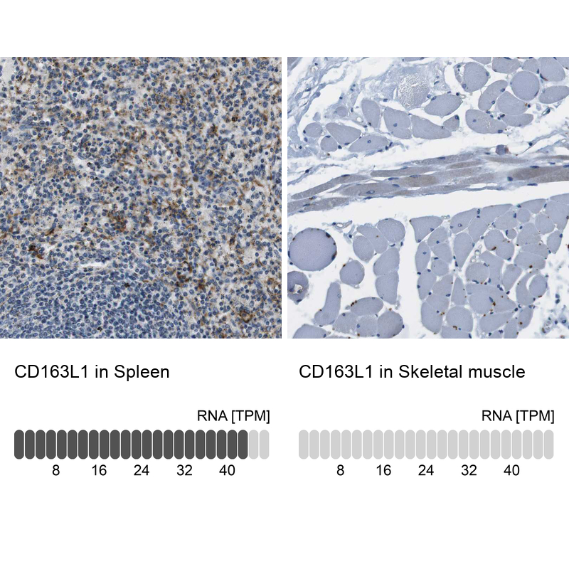
- Main image

- Experimental details
- Immunohistochemistry analysis in human spleen and skeletal muscle tissues using HPA015663 antibody. Corresponding CD163L1 RNA-seq data are presented for the same tissues.
- Sample type
- Human
- Protocol
- Protocol