Explore
 Validate
 Learn
Learn Western blot
 Immunocytochemistry
Antibody data
- Antibody Data
- Antigen structure
- References [1]
- Comments [0]
- Validations
- Immunocytochemistry [1]
- Immunohistochemistry [1]
- Other assay [2]
Submit
Validation data
Reference
Comment
Report error
- Product number
- PA5-32057 - Provider product page

- Provider
- Invitrogen Antibodies
- Product name
- LRRC59 Polyclonal Antibody
- Antibody type
- Polyclonal
- Antigen
- Recombinant full-length protein
- Description
- Recommended positive controls: 293T, A431, HeLa, HepG2. Predicted reactivity: Mouse (98%), Rat (98%), Pig (95%), Bovine (96%). Store product as a concentrated solution. Centrifuge briefly prior to opening the vial.
- Reactivity
- Human, Mouse
- Host
- Rabbit
- Isotype
- IgG
- Vial size
- 100 μL
- Concentration
- 1 mg/mL
- Storage
- Store at 4°C short term. For long term storage, store at -20°C, avoiding freeze/thaw cycles.
Submitted references Translating Proteomic Into Functional Data: An High Mobility Group A1 (HMGA1) Proteomic Signature Has Prognostic Value in Breast Cancer.
Maurizio E, Wiśniewski JR, Ciani Y, Amato A, Arnoldo L, Penzo C, Pegoraro S, Giancotti V, Zambelli A, Piazza S, Manfioletti G, Sgarra R
Molecular & cellular proteomics : MCP 2016 Jan;15(1):109-23
Molecular & cellular proteomics : MCP 2016 Jan;15(1):109-23
No comments: Submit comment
Supportive validation
- Submitted by
- Invitrogen Antibodies (provider)
- Main image
- Experimental details
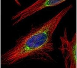
- Immunofluorescent analysis of LRRC59 in methanol-fixed HeLa cells using a LRRC59 polyclonal antibody (Product # PA5-32057) (Green) at a 1:500 dilution. Alpha-tubulin filaments were labeled with Product # PA5-29281 (Red) at a 1:2000.

Supportive validation
- Submitted by
- Invitrogen Antibodies (provider)
- Main image
- Experimental details
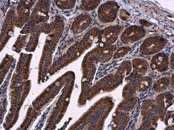
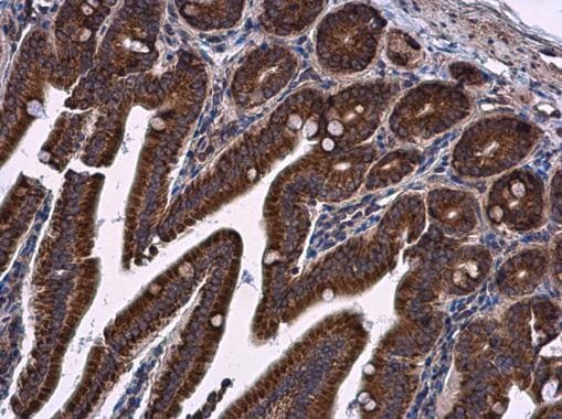
- LRRC59 Polyclonal Antibody detects LRRC59 protein at cytoplasm in mouse duodenum by immunohistochemical analysis. Sample: Paraffin-embedded mouse duodenum. LRRC59 Polyclonal Antibody (Product # PA5-32057) diluted at 1:500. Antigen Retrieval: Citrate buffer, pH 6.0, 15 min.
Supportive validation
- Submitted by
- Invitrogen Antibodies (provider)
- Main image
- Experimental details
- NULL

- Submitted by
- Invitrogen Antibodies (provider)
- Main image
- Experimental details
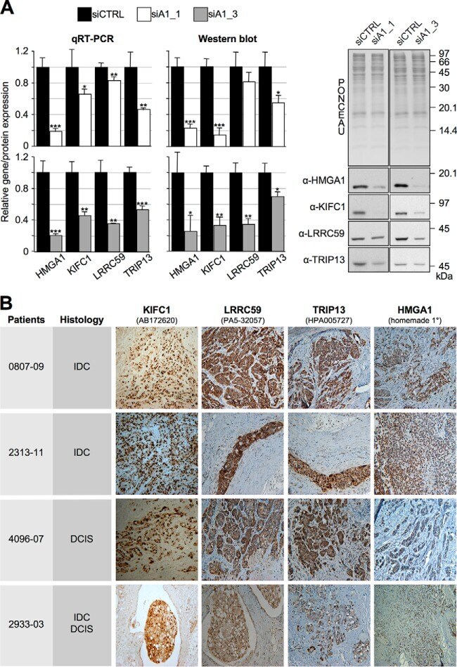
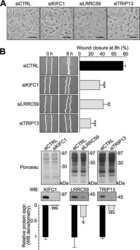
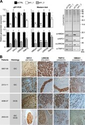
- Fig. 5. The expression of KIFC1, LRRC59, and TRIP13 is linked to HMGA1. MDA-MB-231 cells were treated with control (siCTRL) or HMGA1-targeting siRNAs (siA1_1 and siA1_3) for 72 h. A , mRNA and protein expression levels of the indicated genes were analyzed by qRT-PCR and Western blot. Gene expression levels in HMGA1-silenced cells were compared with that of siCTRL cells. GAPDH was used for normalization. Representative WB analyses are shown together with red ponceau stained membranes to verify total protein normalization. The histogram graphs relative to Western blot analyses were obtained using densitometric analyses (siCTRL versus siA1_1 and siA1_3). The bars indicate the mean +- S.D. ( n = 3). Statistical significance was assessed with Student's t test (*: p < 0.05; **: p < 0.01; ***: p < 0.001). B , Immunohistochemistry analyses performed on breast cancer specimens (IDC, invasive ductal carcinoma; DCIS, ductal carcinoma in situ ; IDC/DCIS, mixed IDC and DCIS) positive for KIFC1, LRRC59, and TRIP13 and HMGA1.