 Explore
 Validate
 Learn
Learn Western blot
 Immunocytochemistry
Antibody data
- Antibody Data
- Antigen structure
- References [3]
- Comments [0]
- Validations
- Immunocytochemistry [1]
Submit
Validation data
Reference
Comment
Report error
- Product number
- HPA041698 - Provider product page

- Provider
- Atlas Antibodies
- Proper citation
- Atlas Antibodies Cat#HPA041698, RRID:AB_10796781
- Product name
- Anti-BLVRB
- Antibody type
- Polyclonal
- Description
- Polyclonal Antibody against Human BLVRB, Gene description: biliverdin reductase B (flavin reductase (NADPH)), Alternative Gene Names: FLR, SDR43U1, Validated applications: WB, IHC, ICC, Uniprot ID: P30043, Storage: Store at +4°C for short term storage. Long time storage is recommended at -20°C.
- Reactivity
- Human
- Host
- Rabbit
- Conjugate
- Unconjugated
- Isotype
- IgG
- Vial size
- 100 µl
- Concentration
- 0.2 mg/ml
- Storage
- Store at +4°C for short term storage. Long time storage is recommended at -20°C.
- Handling
- The antibody solution should be gently mixed before use.
Submitted references Biliverdin Reductase B Is a Plasma Biomarker for Intraplaque Hemorrhage and a Predictor of Ischemic Stroke in Patients with Symptomatic Carotid Atherosclerosis
Biliverdin reductase B impairs cholangiocarcinoma cell motility by inhibiting the Notch/Snail signaling pathway.
Novel Multiomics Profiling of Human Carotid Atherosclerotic Plaques and Plasma Reveals Biliverdin Reductase B as a Marker of Intraplaque Hemorrhage.
Chemaly M, Marlevi D, Iglesias M, Lengquist M, Kronqvist M, Bos D, van Dam-Nolen D, van der Kolk A, Hendrikse J, Kassem M, Matic L, Odeberg J, de Vries M, Kooi M, Hedin U
Biomolecules 2023;13(6):882
Biomolecules 2023;13(6):882
Biliverdin reductase B impairs cholangiocarcinoma cell motility by inhibiting the Notch/Snail signaling pathway.
Gao Z, Ni X, Zheng B, Sun W, Wan W, Liu H, Ni X, Suo T, Li N, Liu H, Shen S
Journal of Cancer 2022;13(7):2159-2170
Journal of Cancer 2022;13(7):2159-2170
Novel Multiomics Profiling of Human Carotid Atherosclerotic Plaques and Plasma Reveals Biliverdin Reductase B as a Marker of Intraplaque Hemorrhage.
Matic LP, Jesus Iglesias M, Vesterlund M, Lengquist M, Hong MG, Saieed S, Sanchez-Rivera L, Berg M, Razuvaev A, Kronqvist M, Lund K, Caidahl K, Gillgren P, Pontén F, Uhlén M, Schwenk JM, Hansson GK, Paulsson-Berne G, Fagman E, Roy J, Hultgren R, Bergström G, Lehtiö J, Odeberg J, Hedin U
JACC. Basic to translational science 2018 Aug;3(4):464-480
JACC. Basic to translational science 2018 Aug;3(4):464-480
No comments: Submit comment
Supportive validation
- Submitted by
- Atlas Antibodies (provider)
- Main image
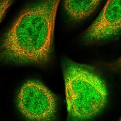
- Experimental details
- Immunofluorescent staining of human cell line U-2 OS shows localization to nucleoplasm & cytosol.
- Sample type
- Human
