Explore
 Validate
 Learn
Learn Flow cytometry
Antibody data
- Antibody Data
- Antigen structure
- References [3]
- Comments [0]
- Validations
- Flow cytometry [2]
- Other assay [4]
Submit
Validation data
Reference
Comment
Report error
- Product number
- 48-9884-42 - Provider product page

- Provider
- Invitrogen Antibodies
- Product name
- CD235a (Glycophorin A) Monoclonal Antibody (6A7M), eFluor™ 450, eBioscience™
- Antibody type
- Monoclonal
- Antigen
- Other
- Description
- Description: The monoclonal antibody 6A7M recognizes human glycophorin A (also known as CD235a). The antibody reacts with the M allele. Glycophorin A is a 151 amino acid sialoglycoprotein found on the erythrocyte (RBC) and erthyroid progenitor cell membrane at about 500,000 copies per cell. The gene for glycophorin resides on chromosome 4 and has 2 allelic forms: M and N, which differ in two amino acids. The M group possesses Ser1 and Gly5 while the N group has Leu1 and Glu5. Recent data suggest that exposure to toxins can cause mutation or loss of an allele resulting in phenotypic changes. Studies are also beginning to correlate genotype/phenotype with predisposition to cancer and heart disease. Applications Reported: This 6A7M antibody has been reported for use in flow cytometric analysis. Applications Tested: This 6A7M antibody has been pre-titrated and tested bu flow cytometric analysis of human peripheral blood cells. This can be used at 5 µL (0.5 µg) per test. A test is defined as the amount (µg) of antibody that will stain a cell sample in a final volume of 100 µL. Cell number should be determined empirically but can range from 10^5 to 10^8 cells/test. eFluor® 450 is an alternative to Pacific Blue®. eFluor® 450 emits at 445 nm and is excited with the Violet laser (405 nm). Please make sure that your instrument is capable of detecting this fluorochome. Excitation: 405 nm; Emission: 445 nm; Laser: Violet Laser. Filtration: 0.2 µm post-manufacturing filtered.
- Reactivity
- Human
- Host
- Mouse
- Isotype
- IgG
- Antibody clone number
- 6A7M
- Vial size
- 100 Tests
- Concentration
- 5 μL/Test
- Storage
- 4°C, store in dark, DO NOT FREEZE!
Submitted references Enforced Expression of HOXB4 in Human Embryonic Stem Cells Enhances the Production of Hematopoietic Progenitors but Has No Effect on the Maturation of Red Blood Cells.
Long-term erythropoiesis from constant numbers of CD34+ cells in serum-free cultures initiated with highly purified progenitor cells from human bone marrow.
Flow cytometric characterization of normal and variant cells with monoclonal antibodies specific for glycophorin A.
Jackson M, Ma R, Taylor AH, Axton RA, Easterbrook J, Kydonaki M, Olivier E, Marenah L, Stanley EG, Elefanty AG, Mountford JC, Forrester LM
Stem cells translational medicine 2016 Aug;5(8):981-90
Stem cells translational medicine 2016 Aug;5(8):981-90
Long-term erythropoiesis from constant numbers of CD34+ cells in serum-free cultures initiated with highly purified progenitor cells from human bone marrow.
Lansdorp PM, Dragowska W
The Journal of experimental medicine 1992 Jun 1;175(6):1501-9
The Journal of experimental medicine 1992 Jun 1;175(6):1501-9
Flow cytometric characterization of normal and variant cells with monoclonal antibodies specific for glycophorin A.
Langlois RG, Bigbee WL, Jensen RH
Journal of immunology (Baltimore, Md. : 1950) 1985 Jun;134(6):4009-17
Journal of immunology (Baltimore, Md. : 1950) 1985 Jun;134(6):4009-17
No comments: Submit comment
Supportive validation
- Submitted by
- Invitrogen Antibodies (provider)
- Main image
- Experimental details
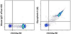
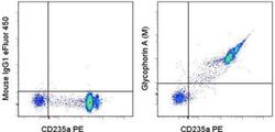
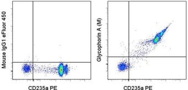
- Staining of normal human peripheral blood cells with Anti-Human CD235a (Glycophorin A) PE (Product # 12-9987-82) and Mouse IgG1 K Isotype Control eFluor® 450 (Product # 48-4714-82) (left) or Anti-Human Glycophorin A (M) eFluor® 450 (right). RBC gate used for analysis.

- Submitted by
- Invitrogen Antibodies (provider)
- Main image
- Experimental details
- Staining of normal human peripheral blood cells with Anti-Human CD235a (Glycophorin A) PE (Product # 12-9987-82) and Mouse IgG1 K Isotype Control eFluor® 450 (Product # 48-4714-82) (left) or Anti-Human Glycophorin A (M) eFluor® 450 (right). RBC gate used for analysis.
Supportive validation
- Submitted by
- Invitrogen Antibodies (provider)
- Main image

- Experimental details
- NULL
- Submitted by
- Invitrogen Antibodies (provider)
- Main image
- Experimental details
- NULL
- Submitted by
- Invitrogen Antibodies (provider)
- Main image

- Experimental details
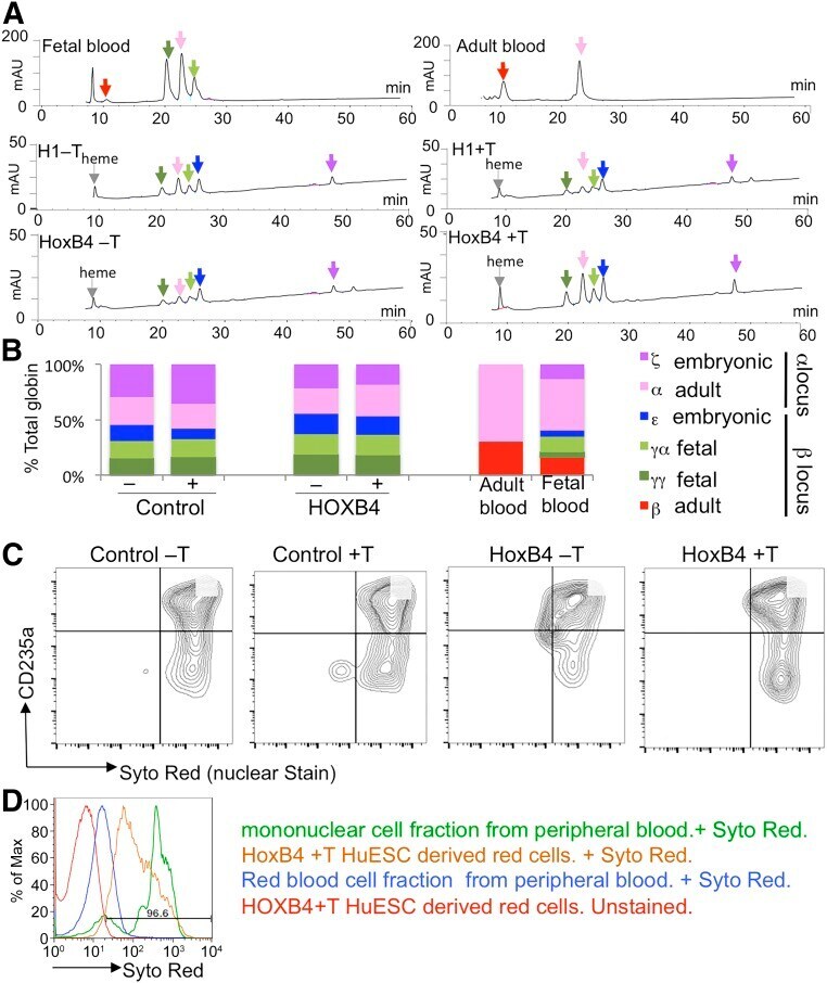
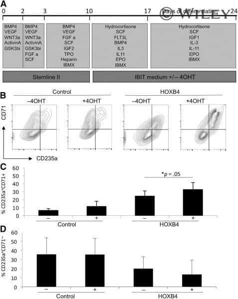
- Figure 4. Activation of HOXB4 resulted in a modest increase in the proportion of immature CD235a + /CD71 + erythroid cells. HOXB4 was activated with 4OHT from days 10 to 24 (A) . Production of erythroid cells was monitored by expression of CD235a and CD71 by flow cytometry at day 24 (B-D) . Data were generated from four independent experiments with error bars representing the SEM. *, p = .05. Abbreviations: 4OHT, tamoxifen; BMP4, bone morphogenetic protein 4; C, cell; CFU, colony-forming unit; EPO, erythropoietin; FGF a, fibroblast growth factor-alpha; FLT3L, FMS-like tyrosine kinase receptor 3 ligand; GSK, glycogen synthase kinase; IBMX, isobutylmethylxanthine; IGF1, IGF2, insulin-like growth factor 1, 2; IL, interleukin; Q, quartile; SCF, stem cell factor; TPO, thrombopoietin; VEGF, vascular endothelial growth factor.

- Submitted by
- Invitrogen Antibodies (provider)
- Main image
- Experimental details
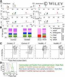
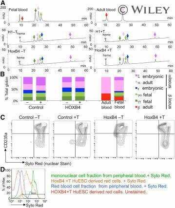
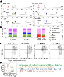
- Figure 5. HOXB4 activation had no significant effect on the maturation of pluripotent stem cell-derived erythroid cells. High-performance liquid chromatography analysis of cell lysates generated at day 24 of the differentiation protocol from control (H1) and HOXB4-activated cells [in the presence (+T) and absence (-T) of tamoxifen] (A) . The amount of the different globins detected as a proportion of the total globin content was calculated in six independent experiments (B) . Adult peripheral blood and fetal blood were included for comparison. Enucleation of control and HOXB4-expressing human embryonic stem cells (hESCs) in the presence (+T) and absence (-T) of tamoxifen was assessed by flow cytometry using the nuclear Syto Red stain and the erythroid marker, CD235a (C) . A representative histogram of SYTO Red staining of control enucleated peripheral blood samples (blue), nucleated mononuclear cells (green), and hESCs after HOXB4 activation (orange) are shown together with the unstained control hESCs (D) . Abbreviation: HuESC, human embryonic stem cell.