Explore
 Validate
 Learn
Learn Western blot
 Immunocytochemistry
Antibody data
- Antibody Data
- Antigen structure
- References [3]
- Comments [0]
- Validations
- Immunocytochemistry [2]
- Other assay [6]
Submit
Validation data
Reference
Comment
Report error
- Product number
- PA5-29140 - Provider product page

- Provider
- Invitrogen Antibodies
- Product name
- TDG Polyclonal Antibody
- Antibody type
- Polyclonal
- Antigen
- Recombinant full-length protein
- Description
- Recommended positive controls: HeLa, Jurkat, Raji, NCI-H929, TDG-transfected 293T. Predicted reactivity: Human (99%), Mouse (88%), Rat (87%), Bovine (91%). Store product as a concentrated solution. Centrifuge briefly prior to opening the vial.
- Reactivity
- Human, Mouse
- Host
- Rabbit
- Isotype
- IgG
- Vial size
- 100 μL
- Concentration
- 1 mg/mL
- Storage
- Store at 4°C short term. For long term storage, store at -20°C, avoiding freeze/thaw cycles.
Submitted references Direct and Base Excision Repair-Mediated Regulation of a GC-Rich cis-Element in Response to 5-Formylcytosine and 5-Carboxycytosine.
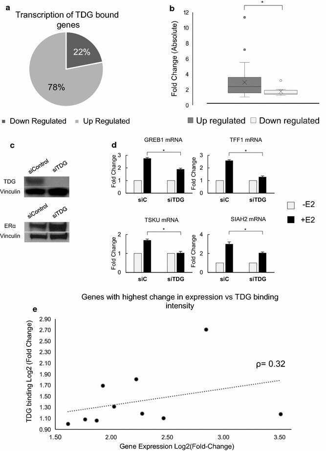
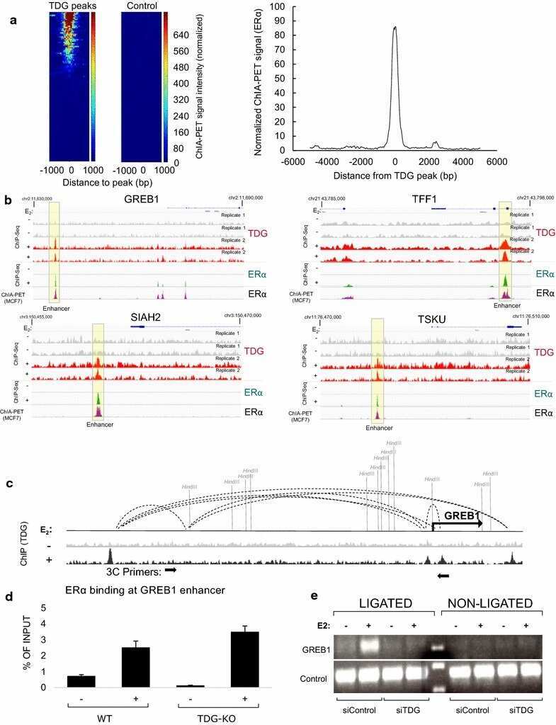
Genome-wide analysis reveals a role for TDG in estrogen receptor-mediated enhancer RNA transcription and 3-dimensional reorganization.
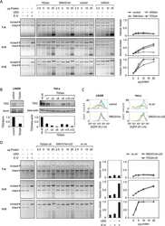
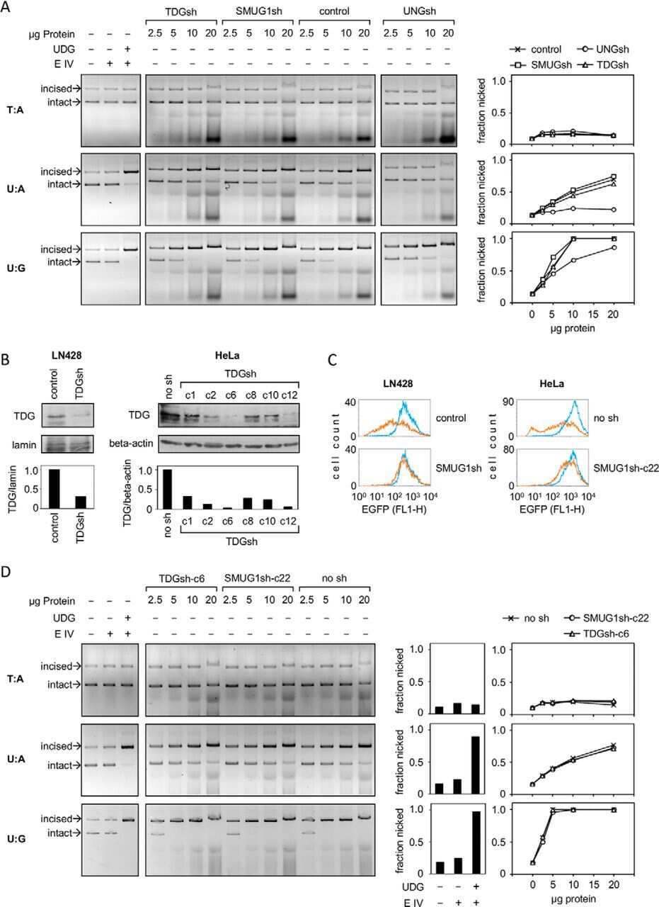
Excision of uracil from transcribed DNA negatively affects gene expression.
Müller N, Ponkkonen E, Carell T, Khobta A
International journal of molecular sciences 2021 Oct 13;22(20)
International journal of molecular sciences 2021 Oct 13;22(20)
Genome-wide analysis reveals a role for TDG in estrogen receptor-mediated enhancer RNA transcription and 3-dimensional reorganization.
Kolendowski B, Hassan H, Krstic M, Isovic M, Thillainadesan G, Chambers AF, Tuck AB, Torchia J
Epigenetics & chromatin 2018 Jan 29;11(1):5
Epigenetics & chromatin 2018 Jan 29;11(1):5
Excision of uracil from transcribed DNA negatively affects gene expression.
Lühnsdorf B, Epe B, Khobta A
The Journal of biological chemistry 2014 Aug 8;289(32):22008-18
The Journal of biological chemistry 2014 Aug 8;289(32):22008-18
No comments: Submit comment
Supportive validation
- Submitted by
- Invitrogen Antibodies (provider)
- Main image

- Experimental details
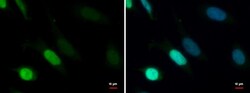
- TDG Polyclonal Antibody detects TDG protein at nucleus by immunofluorescent analysis. Sample: HeLa cells were fixed in 4% paraformaldehyde at RT for 15 min. Green: TDG protein stained by TDG Polyclonal Antibody (Product # PA5-29140) diluted at 1:1,000. Blue: Hoechst 33342 staining.

- Submitted by
- Invitrogen Antibodies (provider)
- Main image
- Experimental details
- TDG Polyclonal Antibody detects TDG protein at nucleus by immunofluorescent analysis. Sample: HeLa cells were fixed in 4% paraformaldehyde at RT for 15 min. Green: TDG protein stained by TDG Polyclonal Antibody (Product # PA5-29140) diluted at 1:1,000. Blue: Hoechst 33342 staining.

Supportive validation
- Submitted by
- Invitrogen Antibodies (provider)
- Main image
- Experimental details
- NULL
- Submitted by
- Invitrogen Antibodies (provider)
- Main image

- Experimental details
- NULL
- Submitted by
- Invitrogen Antibodies (provider)
- Main image

- Experimental details
- NULL
- Submitted by
- Invitrogen Antibodies (provider)
- Main image
- Experimental details
- NULL
- Submitted by
- Invitrogen Antibodies (provider)
- Main image

- Experimental details
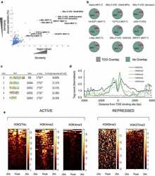
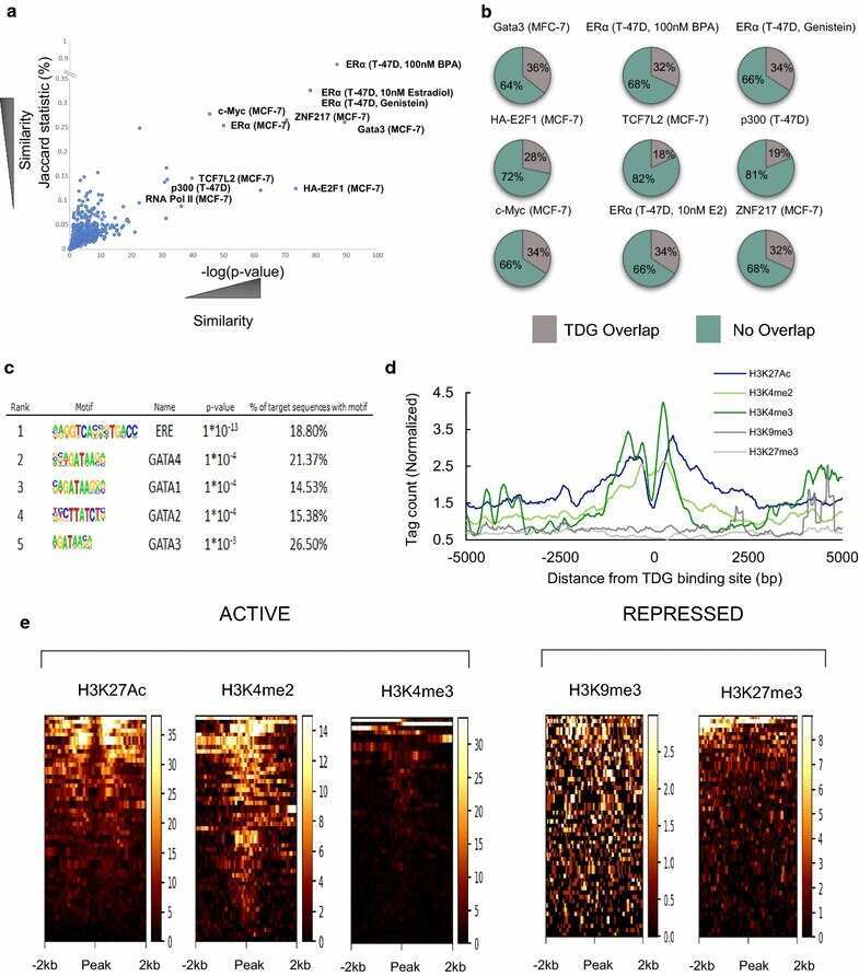
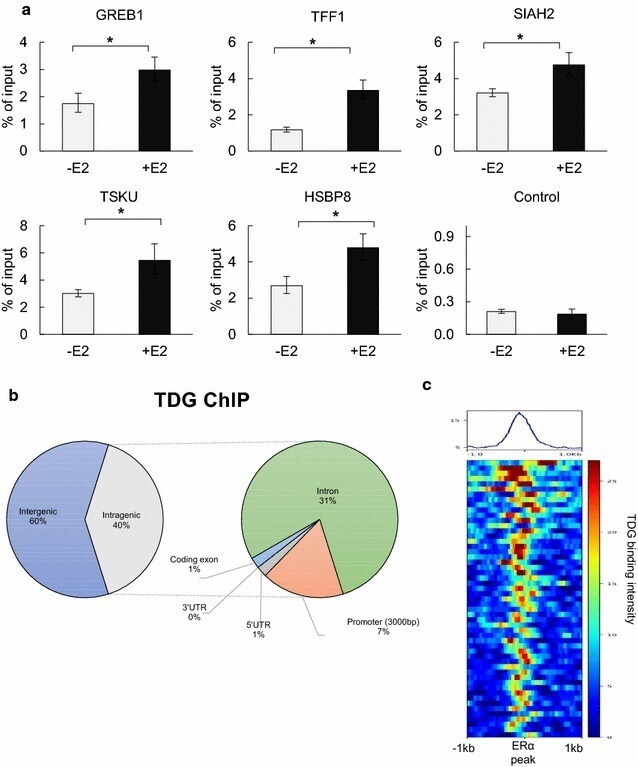
- Fig. 1 Global analysis of E2-dependent TDG localization. a MCF7 cells were treated with 100 nM of E2 (45 min) and ChIP-qPCR was performed using TDG antibody. Region used as negative control shows low level of TDG binding in ChIP-Seq data with no change in levels after E2 treatment. (* p value < 0.05, error bars represent standard deviation of the mean, n > 2). b Sites of E2-dependent TDG binding were mapped to the annotated genome using CEAS. c Sites of ER binding (+/- 1000 bp) overlaid with TDG binding signal, showing strong relationship between location of TDG binding and ER binding at these regions
- Submitted by
- Invitrogen Antibodies (provider)
- Main image
- Experimental details
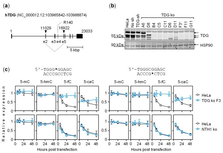
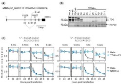
- Figure 5 Effect of TDG knockout on the activity of GC box containing 5-mC/5-hmC/5-fC/5-caC. ( a ) Strategy for TDG knockout in Hela cells. The TDG gene was targeted simultaneously at two different sites by a pair of single guide RNAs enclosing the codon for the catalytic R140 residue. The Cas9 cut sites are indicated by arrowheads. ( b ) Validation of TDG negative clones by Western blotting. Clone F3 further used as a transfection host for the gene expression analyses is marked (*). ( c ) Expression time course of constructs containing 5-mC/5-hmC/5-fC/5-caC in the purine-rich (left group of plots) or the pyrimidine-rich GC box strand (right). Isogenic clonal TDG knockout (upper row) and NTH1 knockout (lower row) cell lines were compared with the parental HeLa cell line (overlaid in plots). All cell lines were transfected in parallel with the same sets of reporter constructs. Results of n = 3 independent experiments (mean +- SD).