Explore
 Validate
 Learn
LearnMA1-016
antibody from Invitrogen Antibodies
Targeting: LIN28A
CSDD1, FLJ12457, LIN-28, LIN28, ZCCHC1
 Western blot
 Immunocytochemistry
Antibody data
- Antibody Data
- Antigen structure
- References [2]
- Comments [0]
- Validations
- Immunocytochemistry [8]
- Immunoprecipitation [1]
- Immunohistochemistry [2]
- Flow cytometry [4]
- Chromatin Immunoprecipitation [2]
- Other assay [1]
Submit
Validation data
Reference
Comment
Report error
- Product number
- MA1-016 - Provider product page

- Provider
- Invitrogen Antibodies
- Product name
- LIN28A Monoclonal Antibody (14E6-4E6)
- Antibody type
- Monoclonal
- Antigen
- Recombinant full-length protein
- Description
- MA1-016 has been successfully used in Western blot, immunofluorescence, ChIP, flow cytometry, IHC (P) and immunoprecipitation applications on human and mouse samples.
- Reactivity
- Human, Mouse
- Host
- Mouse
- Isotype
- IgG
- Antibody clone number
- 14E6-4E6
- Vial size
- 100 μg
- Concentration
- 1 mg/mL
- Storage
- -20°C
Submitted references SUMOylation modulates the LIN28A-let-7 signaling pathway in response to cellular stresses in cancer cells.
Conditionally Stabilized dCas9 Activator for Controlling Gene Expression in Human Cell Reprogramming and Differentiation.
Dou J, Zhang H, Chen R, Shu Z, Yuan H, Zhao X, Wang Y, Huang J, Zhou A, Yu J
Molecular oncology 2020 Sep;14(9):2288-2312
Molecular oncology 2020 Sep;14(9):2288-2312
Conditionally Stabilized dCas9 Activator for Controlling Gene Expression in Human Cell Reprogramming and Differentiation.
Balboa D, Weltner J, Eurola S, Trokovic R, Wartiovaara K, Otonkoski T
Stem cell reports 2015 Sep 8;5(3):448-59
Stem cell reports 2015 Sep 8;5(3):448-59
No comments: Submit comment
Supportive validation
- Submitted by
- Invitrogen Antibodies (provider)
- Main image
- Experimental details
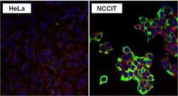
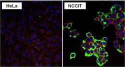
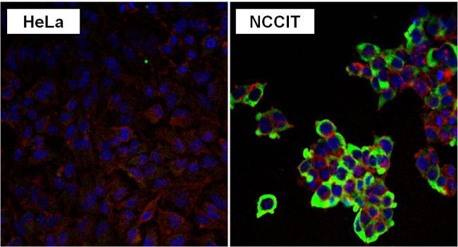
- Immunofluorescent analysis of LIN28 in NCCIT and HeLa cells. Formalin fixed cells were permeabilized with 0.1% Triton X-100 in TBS for 10 minutes at room temperature. Cells were blocked with 1% Blocker BSA (Product # 37525) for 15 minutes at room temperature. Cells were probed with a LIN28 monoclonal antibody (Product # MA1-016) at a dilution of 1:50 for at least 1 hour at room temperature, washed with PBS, and incubated with a DyLight 488-conjugated goat anti-mouse IgG secondary antibody (Product # 35502). F-Actin (red) was stained with DyLight-554 Phalloidin (Product # 21834) and nuclei (blue) were stained with Hoechst 33342 dye (Product # 62249). Images were taken on a Thermo Scientific ArrayScan at 20X magnification.

- Submitted by
- Invitrogen Antibodies (provider)
- Main image

- Experimental details
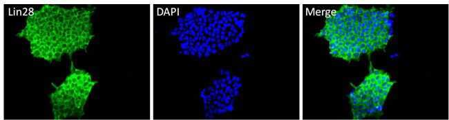
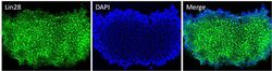
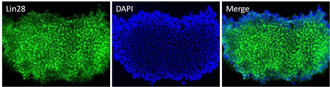
- Immunofluorescent analysis of Lin28 (green) in HEL 11.4 induced IPS cells grown for a few days on Matrigel-coated chamber slides. Cells fixed in 4% paraformaldehyde were permeabilized with 0.1% Triton X-100 for 15 minutes at room temperature. Cells were probed with a Lin28 monoclonal antibody (Product # MA1-016) at a dilution of 1:200 overnight at 4°C, washed with PBST, and incubated with a FITC-conjugated secondary antibody at a dilution of 1:100 for 1 hour at room temperature. Nuclei (blue) were stained with DAPI and cells were analyzed by fluorescence microscopy at 20X magnification.
- Submitted by
- Invitrogen Antibodies (provider)
- Main image

- Experimental details
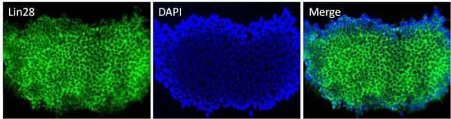
- Immunofluorescent analysis of Lin28 (green) in H9 embryonic stem cells grown for a few days on Matrigel-coated chamber slides. Cells fixed in 4% paraformaldehyde were permeabilized with 0.1% Triton X-100 for 15 minutes at room temperature. Cells were probed with a Lin28 monoclonal antibody (Product # MA1-016) at a dilution of 1:200 overnight at 4°C, washed with PBST, and incubated with a FITC-conjugated secondary antibody at a dilution of 1:100 for 1 hour at room temperature. Nuclei (blue) were stained with DAPI and cells were analyzed by fluorescence microscopy at 20X magnification.
- Submitted by
- Invitrogen Antibodies (provider)
- Main image

- Experimental details
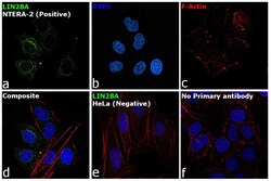
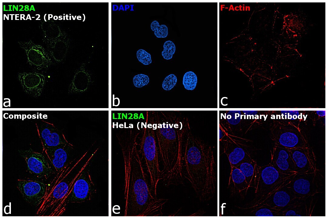
- Immunofluorescence analysis of LIN28A was performed using NTERA-2 and HeLa cells. The cells were fixed with 4% paraformaldehyde for 10 minutes, permeabilized with 0.1% Triton™ X-100 for 15 minutes, and blocked with 2% BSA for 1 hour at room temperature. The cells were labeled with LIN28A Monoclonal Antibody (14E6-4E6) (Product # MA1-016) at 5 µg/mL in 0.1% BSA, incubated at 4 degree celsius overnight and then labeled with Goat anti-Mouse IgG (H+L) Superclonal™ Recombinant Secondary Antibody, Alexa Fluor® 488 (Product # A28175) at a dilution of 1:2000 for 45 minutes at room temperature (Panel a: green). Nuclei (Panel b: blue) were stained with ProLong™ Diamond Antifade Mountant with DAPI (Product # P36962). F-actin (Panel c: red) was stained with Rhodamine Phalloidin (Product # R415, 1:300). Panel d represents the merged image showing cytoplasmic expression of LIN28A in NTERA-2. Panel e represents HeLa cells, showing lesser expression of LIN28A. Panel f represents control NTERA-2 cells with no primary antibody to assess background. The images were captured at 60X magnification.

- Submitted by
- Invitrogen Antibodies (provider)
- Main image
- Experimental details
- Immunofluorescent analysis of LIN28 in NCCIT and HeLa cells. Formalin fixed cells were permeabilized with 0.1% Triton X-100 in TBS for 10 minutes at room temperature. Cells were blocked with 1% Blocker BSA (Product # 37525) for 15 minutes at room temperature. Cells were probed with a LIN28 monoclonal antibody (Product # MA1-016) at a dilution of 1:50 for at least 1 hour at room temperature, washed with PBS, and incubated with a DyLight 488-conjugated goat anti-mouse IgG secondary antibody (Product # 35502). F-Actin (red) was stained with DyLight-554 Phalloidin (Product # 21834) and nuclei (blue) were stained with Hoechst 33342 dye (Product # 62249). Images were taken on a Thermo Scientific ArrayScan at 20X magnification.
- Submitted by
- Invitrogen Antibodies (provider)
- Main image

- Experimental details
- Immunofluorescent analysis of Lin28 (green) in HEL 11.4 induced IPS cells grown for a few days on Matrigel-coated chamber slides. Cells fixed in 4% paraformaldehyde were permeabilized with 0.1% Triton X-100 for 15 minutes at room temperature. Cells were probed with a Lin28 monoclonal antibody (Product # MA1-016) at a dilution of 1:200 overnight at 4°C, washed with PBST, and incubated with a FITC-conjugated secondary antibody at a dilution of 1:100 for 1 hour at room temperature. Nuclei (blue) were stained with DAPI and cells were analyzed by fluorescence microscopy at 20X magnification.

- Submitted by
- Invitrogen Antibodies (provider)
- Main image
- Experimental details
- Immunofluorescent analysis of Lin28 (green) in H9 embryonic stem cells grown for a few days on Matrigel-coated chamber slides. Cells fixed in 4% paraformaldehyde were permeabilized with 0.1% Triton X-100 for 15 minutes at room temperature. Cells were probed with a Lin28 monoclonal antibody (Product # MA1-016) at a dilution of 1:200 overnight at 4°C, washed with PBST, and incubated with a FITC-conjugated secondary antibody at a dilution of 1:100 for 1 hour at room temperature. Nuclei (blue) were stained with DAPI and cells were analyzed by fluorescence microscopy at 20X magnification.
- Submitted by
- Invitrogen Antibodies (provider)
- Main image
- Experimental details
- Immunofluorescence analysis of LIN28A was performed using NTERA-2 and HeLa cells. The cells were fixed with 4% paraformaldehyde for 10 minutes, permeabilized with 0.1% Triton™ X-100 for 15 minutes, and blocked with 2% BSA for 1 hour at room temperature. The cells were labeled with LIN28A Monoclonal Antibody (14E6-4E6) (Product # MA1-016) at 5 µg/mL in 0.1% BSA, incubated at 4 degree celsius overnight and then labeled with Goat anti-Mouse IgG (H+L) Superclonal™ Recombinant Secondary Antibody, Alexa Fluor® 488 (Product # A28175) at a dilution of 1:2000 for 45 minutes at room temperature (Panel a: green). Nuclei (Panel b: blue) were stained with ProLong™ Diamond Antifade Mountant with DAPI (Product # P36962). F-actin (Panel c: red) was stained with Rhodamine Phalloidin (Product # R415, 1:300). Panel d represents the merged image showing cytoplasmic expression of LIN28A in NTERA-2. Panel e represents HeLa cells, showing lesser expression of LIN28A. Panel f represents control NTERA-2 cells with no primary antibody to assess background. The images were captured at 60X magnification.
Supportive validation
- Submitted by
- Invitrogen Antibodies (provider)
- Main image

- Experimental details
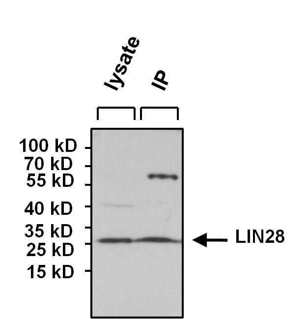
- Immunoprecipitation of LIN28 was performed using NCCIT whole cell lysate. Antigen-antibody complexes were formed by incubating 700 µg of lysate with 5 µg of an LIN28 monoclonal antibody (Product # MA1-016) overnight on a rocking platform at 4°C. The immune complexes were captured on 50 µl Protein A/G Agarose (Product # 20421), washed extensively, and eluted with 5X Lane Marker Reducing Sample Buffer (Product # 39000). NCCIT whole cell lysate (70 µg) was loaded as a positive control for detection. Samples were resolved on a 4-20% Tris-HCl polyacrylamide gel, transferred to a PVDF membrane, and blocked with 5% BSA/TBS-0.1%Tween for at least 1 hour. The membrane was probed with a LIN28 monoclonal antibody (Product # MA1-016) at a dilution of 1:1000 overnight rotating at 4°C, washed in TBST, and probed with Clean-blot IP Detection Reagent (Product # 21230) at a dilution of 1:1000 for at least 1 hour. Chemiluminescent detection was performed using SuperSignal West Dura (Product # 34075).

Supportive validation
- Submitted by
- Invitrogen Antibodies (provider)
- Main image
- Experimental details
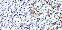
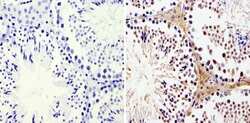
- Immunohistochemistry analysis of LIN28 showing staining in the cytoplasm of paraffin-embedded human seminoma (right) compared with a negative control without primary antibody (left). To expose target proteins, antigen retrieval was performed using 10mM sodium citrate (pH 6.0), microwaved for 8-15 min. Following antigen retrieval, tissues were blocked in 3% H2O2-methanol for 15 min at room temperature, washed with ddH2O and PBS, and then probed with a LIN28 monoclonal antibody (Product # MA1-016) diluted in 3% BSA-PBS at a dilution of 1:200 overnight at 4°C in a humidified chamber. Tissues were washed extensively in PBST and detection was performed using an HRP-conjugated secondary antibody followed by colorimetric detection using a DAB kit. Tissues were counterstained with hematoxylin and dehydrated with ethanol and xylene to prep for mounting.
- Submitted by
- Invitrogen Antibodies (provider)
- Main image
- Experimental details
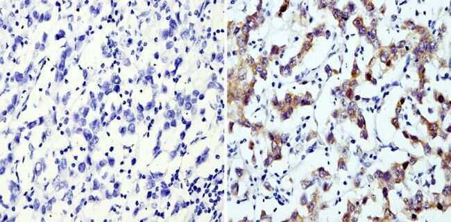
- Immunohistochemistry analysis of LIN28 showing staining in the nucleus and cytoplasm of paraffin-embedded mouse testis tissue (right) compared with a negative control without primary antibody (left). To expose target proteins, antigen retrieval was performed using 10mM sodium citrate (pH 6.0), microwaved for 8-15 min. Following antigen retrieval, tissues were blocked in 3% H2O2-methanol for 15 min at room temperature, washed with ddH2O and PBS, and then probed with a LIN28 monoclonal antibody (Product # MA1-016) diluted in 3% BSA-PBS at a dilution of 1:20 overnight at 4°C in a humidified chamber. Tissues were washed extensively in PBST and detection was performed using an HRP-conjugated secondary antibody followed by colorimetric detection using a DAB kit. Tissues were counterstained with hematoxylin and dehydrated with ethanol and xylene to prep for mounting.

Supportive validation
- Submitted by
- Invitrogen Antibodies (provider)
- Main image
- Experimental details
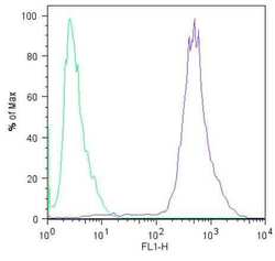
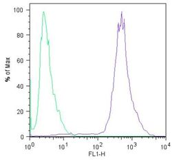
- Flow cytometric analysis of Lin28 (blue histogram) on HEL 11.4 induced IPS cells. To generate single cells suspensions, colonies were treated with TrypLE cell dissociation enzyme for 5 minutes at 37°C. Cells were incubated with a Lin28 monoclonal antibody (Product # MA1-016) or mouse IgG (green histogram) at a dilution of 1:100 for 1 hour on ice, washed with PBS + 5% fetal calf serum (FACS buffer), and incubated with a FITC-conjugated secondary antibody at a dilution of 1:200 for 30 minutes on ice. Cells were washed with cold FACS buffer, resuspended in 500 µL of FACS buffer containing 10 µL of 4% paraformaldehyde, and analyzed on a flow cytometer.

- Submitted by
- Invitrogen Antibodies (provider)
- Main image
- Experimental details
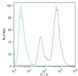
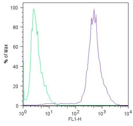
- Flow cytometric analysis of Lin28 (blue histogram) on H9 embryonic stem cells. To generate single cells suspensions, colonies were treated with TrypLE cell dissociation enzyme for 5 minutes at 37°C. Cells were incubated with a Lin28 monoclonal antibody (Product # MA1-016) or mouse IgG (green histogram) at a dilution of 1:100 for 1 hour on ice, washed with PBS + 5% fetal calf serum (FACS buffer), and incubated with a FITC-conjugated secondary antibody at a dilution of 1:200 for 30 minutes on ice. Cells were washed with cold FACS buffer, resuspended in 500 µL of FACS buffer containing 10 µL of 4% paraformaldehyde, and analyzed on a flow cytometer.
- Submitted by
- Invitrogen Antibodies (provider)
- Main image
- Experimental details
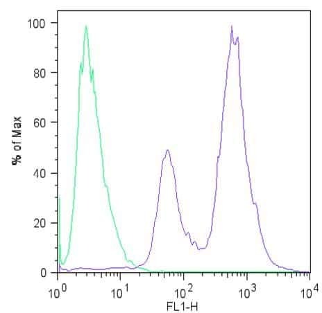
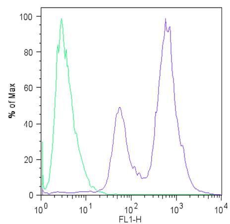
- Flow cytometric analysis of Lin28 (blue histogram) on HEL 11.4 induced IPS cells. To generate single cells suspensions, colonies were treated with TrypLE cell dissociation enzyme for 5 minutes at 37°C. Cells were incubated with a Lin28 monoclonal antibody (Product # MA1-016) or mouse IgG (green histogram) at a dilution of 1:100 for 1 hour on ice, washed with PBS + 5% fetal calf serum (FACS buffer), and incubated with a FITC-conjugated secondary antibody at a dilution of 1:200 for 30 minutes on ice. Cells were washed with cold FACS buffer, resuspended in 500 µL of FACS buffer containing 10 µL of 4% paraformaldehyde, and analyzed on a flow cytometer.
- Submitted by
- Invitrogen Antibodies (provider)
- Main image

- Experimental details
- Flow cytometric analysis of Lin28 (blue histogram) on H9 embryonic stem cells. To generate single cells suspensions, colonies were treated with TrypLE cell dissociation enzyme for 5 minutes at 37°C. Cells were incubated with a Lin28 monoclonal antibody (Product # MA1-016) or mouse IgG (green histogram) at a dilution of 1:100 for 1 hour on ice, washed with PBS + 5% fetal calf serum (FACS buffer), and incubated with a FITC-conjugated secondary antibody at a dilution of 1:200 for 30 minutes on ice. Cells were washed with cold FACS buffer, resuspended in 500 µL of FACS buffer containing 10 µL of 4% paraformaldehyde, and analyzed on a flow cytometer.
Supportive validation
- Submitted by
- Invitrogen Antibodies (provider)
- Main image
- Experimental details
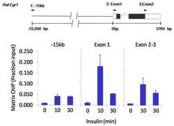
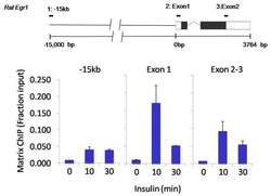
- Chromatin immunoprecipitation analysis of LIN28 was performed using cross-linked chromatin from 1x10^6 HTC-IR rat hepatoma cells treated with insulin for 0, 10, and 30 minutes. Immunoprecipitation was performed using a multiplex microplate Matrix ChIP assay (see reference for Matrix ChIP protocol: http://www.ncbi.nlm.nih.gov/pubmed/22098709) with 1.0 µL/100 µL well volume of a LIN28 monoclonal antibody (Product # MA1-016). Chromatin aliquots from ~1x10^5 cells were used per ChIP pull-down. Quantitative PCR data were done in quadruplicate using 1 µL of eluted DNA in 2 µL SYBR real-time PCR reactions containing primers to amplify -15kb upstream of the Egr1 gene or exon-1 or exon-2-3 of Egr1. PCR calibration curves were generated for each primer pair from a dilution series of sheared total genomic DNA. Quantitation of immunoprecipitated chromatin is presented as signal relative to the total amount of input chromatin. Results represent the mean +/- SEM for three experiments. A schematic representation of the rat Egr-1 locus is shown above the data where boxes represent exons (black boxes = translated regions, white boxes = untranslated regions), the zigzag line represents an intron, and the straight line represents upstream sequence. Regions amplified by Egr-1 primers are represented by black bars. Data courtesy of the Innovators Program.

- Submitted by
- Invitrogen Antibodies (provider)
- Main image
- Experimental details
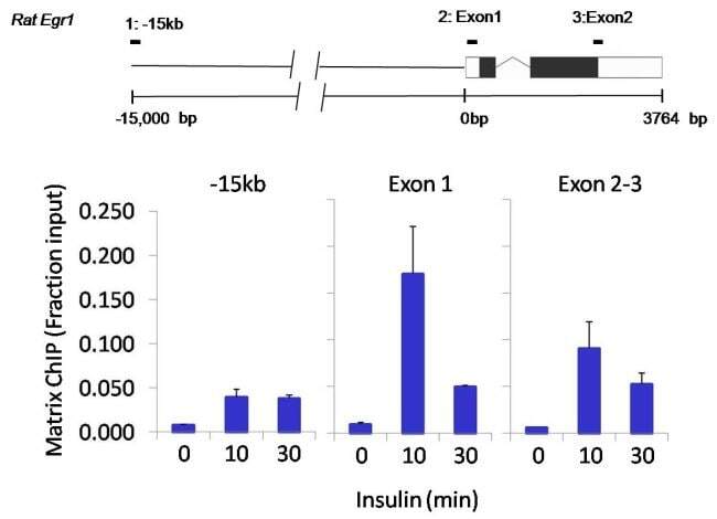
- Chromatin immunoprecipitation analysis of LIN28 was performed using cross-linked chromatin from 1x10^6 HTC-IR rat hepatoma cells treated with insulin for 0, 10, and 30 minutes. Immunoprecipitation was performed using a multiplex microplate Matrix ChIP assay (see reference for Matrix ChIP protocol: http://www.ncbi.nlm.nih.gov/pubmed/22098709) with 1.0 µL/100 µL well volume of a LIN28 monoclonal antibody (Product # MA1-016). Chromatin aliquots from ~1x10^5 cells were used per ChIP pull-down. Quantitative PCR data were done in quadruplicate using 1 µL of eluted DNA in 2 µL SYBR real-time PCR reactions containing primers to amplify -15kb upstream of the Egr1 gene or exon-1 or exon-2-3 of Egr1. PCR calibration curves were generated for each primer pair from a dilution series of sheared total genomic DNA. Quantitation of immunoprecipitated chromatin is presented as signal relative to the total amount of input chromatin. Results represent the mean +/- SEM for three experiments. A schematic representation of the rat Egr-1 locus is shown above the data where boxes represent exons (black boxes = translated regions, white boxes = untranslated regions), the zigzag line represents an intron, and the straight line represents upstream sequence. Regions amplified by Egr-1 primers are represented by black bars. Data courtesy of the Innovators Program.
Supportive validation
- Submitted by
- Invitrogen Antibodies (provider)
- Main image

- Experimental details
- Immunoprecipitation of LIN28 was performed using NCCIT whole cell lysate. Antigen-antibody complexes were formed by incubating 700 µg of lysate with 5 µg of an LIN28 monoclonal antibody (Product # MA1-016) overnight on a rocking platform at 4øC. The immune complexes were captured on 50 µl Protein A/G Agarose (Product # 20421), washed extensively, and eluted with 5X Lane Marker Reducing Sample Buffer (Product # 39000). NCCIT whole cell lysate (70 µg) was loaded as a positive control for detection. Samples were resolved on a 4-20% Tris-HCl polyacrylamide gel, transferred to a PVDF membrane, and blocked with 5% BSA/TBS-0.1%Tween for at least 1 hour. The membrane was probed with a LIN28 monoclonal antibody (Product # MA1-016) at a dilution of 1:1000 overnight rotating at 4øC, washed in TBST, and probed with Clean-blot IP Detection Reagent (Product # 21230) at a dilution of 1:1000 for at least 1 hour. Chemiluminescent detection was performed using SuperSignal West Dura (Product # 34075).