Explore
 Validate
 Learn
LearnMA5-14582
antibody from Invitrogen Antibodies
Targeting: ANO1
DOG1, FLJ10261, ORAOV2, TAOS2, TMEM16A
 Western blot
Antibody data
- Antibody Data
- Antigen structure
- References [5]
- Comments [0]
- Validations
- Western blot [1]
- Immunocytochemistry [2]
- Immunohistochemistry [2]
- Flow cytometry [1]
Submit
Validation data
Reference
Comment
Report error
- Product number
- MA5-14582 - Provider product page

- Provider
- Invitrogen Antibodies
- Product name
- DOG-1 Recombinant Rabbit Monoclonal Antibody (SP31)
- Antibody type
- Monoclonal
- Antigen
- Synthetic peptide
- Description
- MA5-14582 targets Dog1 in IHC (P) applications and shows reactivity with Human samples. The MA5-14582 immunogen is synthetic peptide of human Dog1.
- Reactivity
- Human
- Host
- Rabbit
- Isotype
- IgG
- Antibody clone number
- SP31
- Vial size
- 1 mL
- Concentration
- 0.057 mg/mL
- Storage
- -20°C, Avoid Freeze/Thaw Cycles
Submitted references TMEM16A/ANO1 is differentially expressed in HPV-negative versus HPV-positive head and neck squamous cell carcinoma through promoter methylation.
Plexiform fibromyxoma (plexiform angiomyxoid myofibroblastic tumor) of stomach: an unusual presentation as a fistulating abscess.
Na+/Ca2+ exchangers regulate the migration and proliferation of human gastric myofibroblasts.
Contribution of DOG1 expression to the diagnosis of gastrointestinal stromal tumors.
The KIT Exon 11 Stop Codon Mutation in Gastrointestinal Stromal Tumors: What Is the Clinical Meaning?
Dixit R, Kemp C, Kulich S, Seethala R, Chiosea S, Ling S, Ha PK, Duvvuri U
Scientific reports 2015 Nov 13;5:16657
Scientific reports 2015 Nov 13;5:16657
Plexiform fibromyxoma (plexiform angiomyxoid myofibroblastic tumor) of stomach: an unusual presentation as a fistulating abscess.
Lee PW, Yau DT, Lau PP, Chan JK
International journal of surgical pathology 2014 May;22(3):286-90
International journal of surgical pathology 2014 May;22(3):286-90
Na+/Ca2+ exchangers regulate the migration and proliferation of human gastric myofibroblasts.
Kemény LV, Schnúr A, Czepán M, Rakonczay Z Jr, Gál E, Lonovics J, Lázár G, Simonka Z, Venglovecz V, Maléth J, Judák L, Németh IB, Szabó K, Almássy J, Virág L, Geisz A, Tiszlavicz L, Yule DI, Wittmann T, Varró A, Hegyi P
American journal of physiology. Gastrointestinal and liver physiology 2013 Oct 15;305(8):G552-63
American journal of physiology. Gastrointestinal and liver physiology 2013 Oct 15;305(8):G552-63
Contribution of DOG1 expression to the diagnosis of gastrointestinal stromal tumors.
Kara T, Serinsoz E, Arpaci RB, Gubur O, Orekici G, Ata A, Colak T, Arican A
Pathology, research and practice 2013 Jul;209(7):413-7
Pathology, research and practice 2013 Jul;209(7):413-7
The KIT Exon 11 Stop Codon Mutation in Gastrointestinal Stromal Tumors: What Is the Clinical Meaning?
Michelucci A, Chiappetta C, Cacciotti J, Veccia N, Astri E, Leopizzi M, Prosperi Porta R, Petrozza V, Della Rocca C, Bevilacqua G, Cavazzana A, Di Cristofano C
Gut and liver 2013 Jan;7(1):35-40
Gut and liver 2013 Jan;7(1):35-40
No comments: Submit comment
Supportive validation
- Submitted by
- Invitrogen Antibodies (provider)
- Main image

- Experimental details
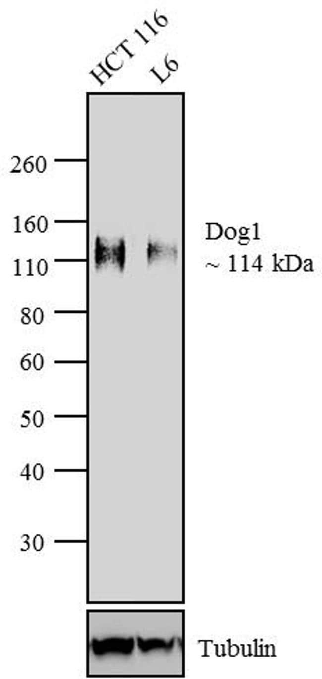
- Western blot analysis of Dog1 was performed using whole cell lysates of HCT 116 (Lane 1) and L6 (Lane 2). The blot was probed with Anti- Dog1 Rabbit monoclonal Antibody (Product # MA5-14582, 1:250 dilution) and detected by chemiluminescence using Goat anti-Rabbit IgG (Heavy Chain) Superclonal™ Secondary Antibody, HRP conjugate (Product # A27036, 0.4 µg/mL, 1:2500 dilution). A 114 kDa band corresponding to Dog1 was observed across cell lines tested. Known quantity of protein samples were electrophoresed using Novex® NuPAGE® 4-12 % Bis-Tris gel (Product # NP0321BOX), XCell SureLock™ Electrophoresis System (Product # EI0002) and Novex® Sharp Pre-Stained Protein Standard (Product # LC5800). Resolved proteins were then transferred onto a nitrocellulose membrane by overnight transfer method. The membrane was probed with the relevant primary and secondary Antibody using iBind™ Flex Western Starter Kit (Product # SLF2000S). Chemiluminescent detection was performed using Pierce™ ECL Western Blotting Substrate (Product # 32106).
Supportive validation
- Submitted by
- Invitrogen Antibodies (provider)
- Main image
- Experimental details
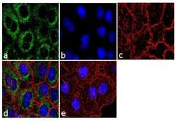
- Immunofluorescence analysis of Dog1 was performed using 70% confluent log phase A549 cells. The cells were fixed with 4% paraformaldehyde for 10 minutes, permeabilized with 0.1% Triton™ X-100 for 10 minutes, and blocked with 1% BSA for 1 hour at room temperature. The cells were labeled with Dog1 (SP31) Rabbit Monoclonal Antibody (Product # MA5-14582) at 1:250 dilution in 0.1% BSA and incubated for 3 hours at room temperature and then labeled with Goat anti-Rabbit IgG (H+L) Superclonal™ Secondary Antibody, Alexa Fluor® 488 conjugate (Product # A27034) a dilution of 1:2000 for 45 minutes at room temperature (Panel a: green). Nuclei (Panel b: blue) were stained with SlowFade® Gold Antifade Mountant with DAPI (Product # S36938). F-actin (Panel c: red) was stained with Rhodamine Phalloidin (Product # R415, 1:300). Panel d represents the merged image showing punctated cytoplasmic localization. Panel e shows the no primary antibody control. The images were captured at 60X magnification.

- Submitted by
- Invitrogen Antibodies (provider)
- Main image

- Experimental details
- Immunofluorescence analysis of Dog1 was performed using 70% confluent log phase A549 cells. The cells were fixed with 4% paraformaldehyde for 10 minutes, permeabilized with 0.1% Triton™ X-100 for 10 minutes, and blocked with 1% BSA for 1 hour at room temperature. The cells were labeled with Dog1 (SP31) Rabbit Monoclonal Antibody (Product # MA5-14582) at 1:250 dilution in 0.1% BSA and incubated for 3 hours at room temperature and then labeled with Goat anti-Rabbit IgG (Heavy Chain) Superclonal™ Secondary Antibody, Alexa Fluor® 488 conjugate (Product # A27034) a dilution of 1:2000 for 45 minutes at room temperature (Panel a: green). Nuclei (Panel b: blue) were stained with SlowFade® Gold Antifade Mountant with DAPI (Product # S36938). F-actin (Panel c: red) was stained with Rhodamine Phalloidin (Product # R415, 1:300). Panel d represents the merged image showing punctated cytoplasmic localization. Panel e shows the no primary antibody control. The images were captured at 60X magnification.

Supportive validation
- Submitted by
- Invitrogen Antibodies (provider)
- Main image

- Experimental details
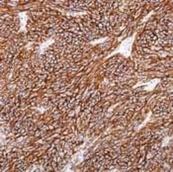
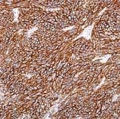
- Formalin-fixed, paraffin-embedded human gastrointestinal stromal tumor stained with rabbit monoclonal DOG1 using peroxidase-conjugate and DAB chromogen. Note cytoplasmic/ membranous staining.

- Submitted by
- Invitrogen Antibodies (provider)
- Main image
- Experimental details
- Formalin-fixed, paraffin-embedded human gastrointestinal stromal tumor stained with rabbit monoclonal DOG1 using peroxidase-conjugate and DAB chromogen. Note cytoplasmic/ membranous staining.
Supportive validation
- Submitted by
- Invitrogen Antibodies (provider)
- Main image
- Experimental details
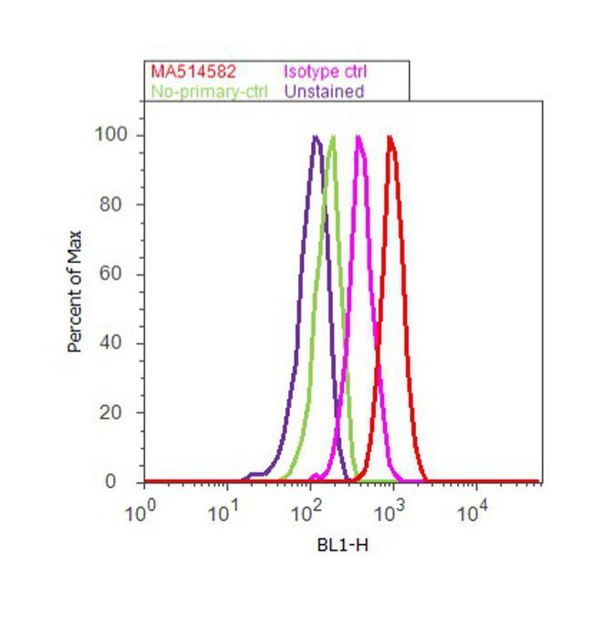
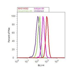
- Flow cytometry analysis of Dog1 was performed using A549 cells. Cells were fixed with 70% ethanol for 10 minutes, permeabilized with 0.25% Triton™ X-100 for 20 minutes, and blocked with 5% BSA for 30 minutes at room temperature. Cells were labeled with Dog1 Rabbit Monoclonal Antibody (MA5-14582, red histogram) or with rabbit isotype control (pink histogram) at 3-5 ug/million cells in 2.5% BSA. After incubation at room temperature for 2 hours, the cells were labeled with Alexa Fluor® 488 Goat Anti-Rabbit Secondary Antibody (A11008) at a dilution of 1:400 for 30 minutes at room temperature. The representative 10,000 cells were acquired and analyzed for each sample using an Attune® Acoustic Focusing Cytometer. The purple histogram represents unstained control cells and the green histogram represents no-primary-antibody control..