 Explore
 Validate
 Learn
Learn Western blot
Antibody data
- Antibody Data
- Antigen structure
- References [0]
- Comments [0]
- Validations
- Western blot [6]
- Immunocytochemistry [4]
- Immunohistochemistry [1]
Submit
Validation data
Reference
Comment
Report error
- Product number
- ARG54707 - Provider product page

- Provider
- Arigo
- Product name
- anti-LC3B antibody
- Antibody type
- Polyclonal
- Antigen
- KLH-conjugated synthetic peptide around aa. 1-30 (N-terminus) of Human LC3 protein (NP_073729.1).
- Description
- This antibody is prepared by Saturated Ammonium Sulfate (SAS) precipitation followed by dialysis against PBS.
- Reactivity
- Human, Mouse
- Host
- Rabbit
- Vial size
- 200 µl
- Storage
- For continuous use, store undiluted antibody at 2-8°C for up to 6 months. For long-term storage, aliquot and store at -20°C. Storage in frost free freezers is not recommended. Avoid repeated freeze/thaw cycles. The antibody solution should be gently mixed before use.
- Handling
- The antibody solution should be gently mixed before use.
No comments: Submit comment
Supportive validation
- Submitted by
- Arigo (provider)
- Main image
- Experimental details
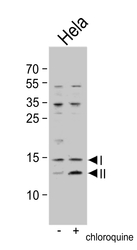
- Western blot: untreated or treated HeLa cell lysate stained with ARG54707 anti-LC3B antibody. Both non-lipidated (arrow, I) and lipidated LC3 (APG8b) (arrow, II) were detected. But pro-LC3 (APG8b) and non-lipidated LC3 (APG8b) were detected in soluble fraction (S).

- Submitted by
- Arigo (provider)
- Main image
- Experimental details
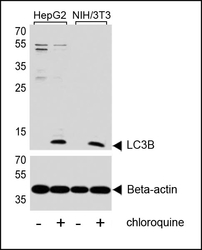
- Western blot: lysates from HepG2, mouse NIH/3T3 cell line, untreated or treated with 50uM chloroquine, stained with ARG54707 anti-LC3B antibody (upper) or Beta-actin (lower).

- Submitted by
- Arigo (provider)
- Main image
- Experimental details
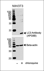
- Western blot: NIH/3T3 cell lysates untreated or treated with chloroquine, stained with ARG54707 anti-LC3B antibody (upper) or Beta-actin (lower).

- Submitted by
- Arigo (provider)
- Main image
- Experimental details
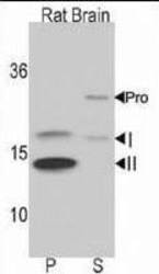
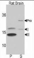
- Western blot: rat brain lysate stained with ARG54707 anti-LC3B antibody. Both non-lipidated (arrow, I) and lipidated LC3 (APG8b) (arrow, II) were detected in membrane fraction (P) but pro-LC3 (APG8b) and non-lipidated LC3 ((APG8b) were detected in soluble fraction (S).
- Submitted by
- Arigo (provider)
- Main image
- Experimental details
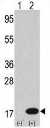
- Western blot: 2 µg of 293 cell line lysates transiently transfected with the LC3 (APG8b) gene stained with ARG54707 anti-LC3B antibody.

- Submitted by
- Arigo (provider)
- Main image
- Experimental details
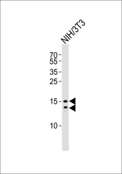
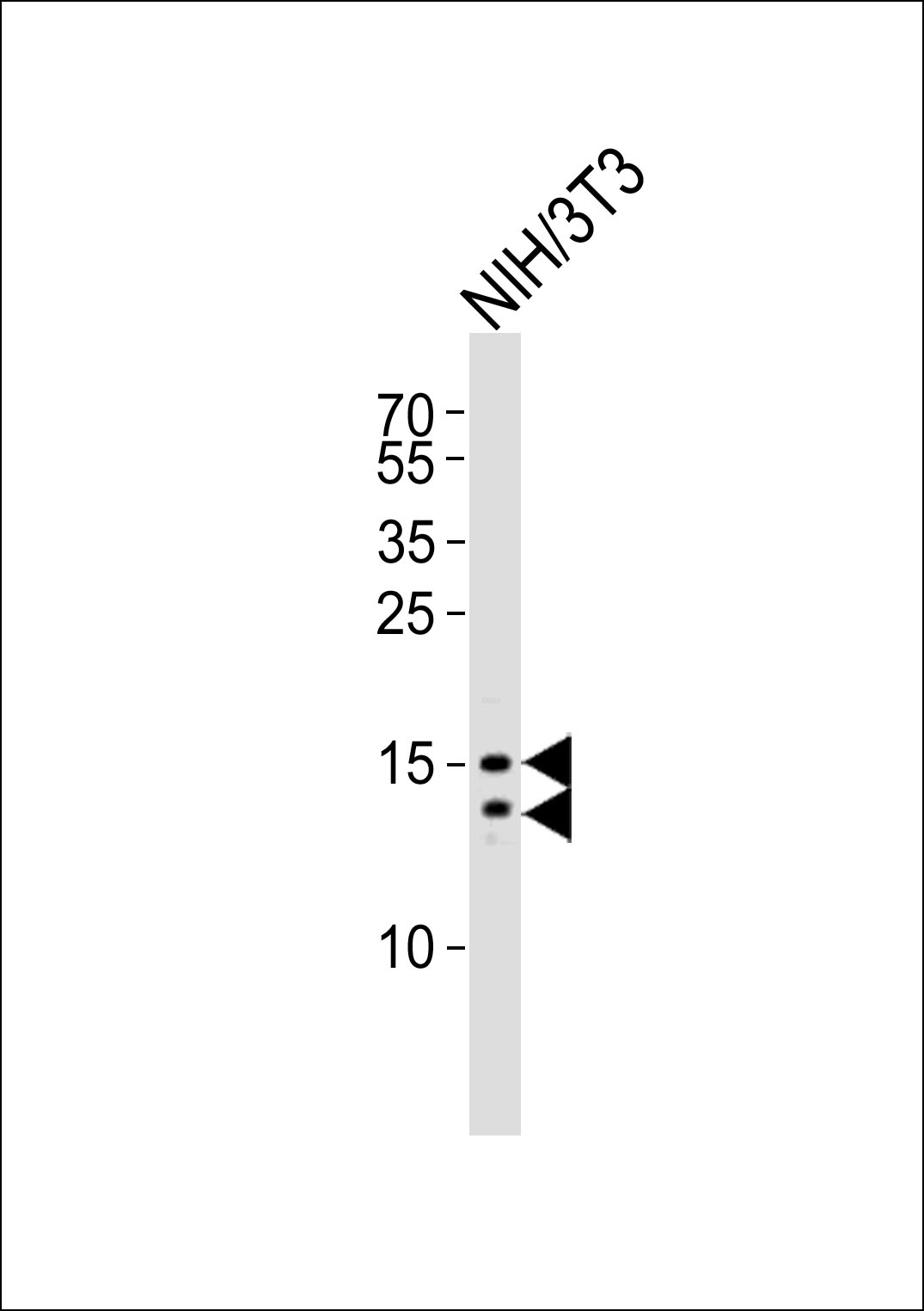
- Western blot: 20 µg of mouse NIH/3T3 cell lysate stained with ARG54707 anti-LC3B antibody at 1:1000 dilution.
Supportive validation
- Submitted by
- Arigo (provider)
- Main image
- Experimental details
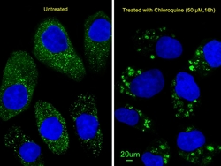
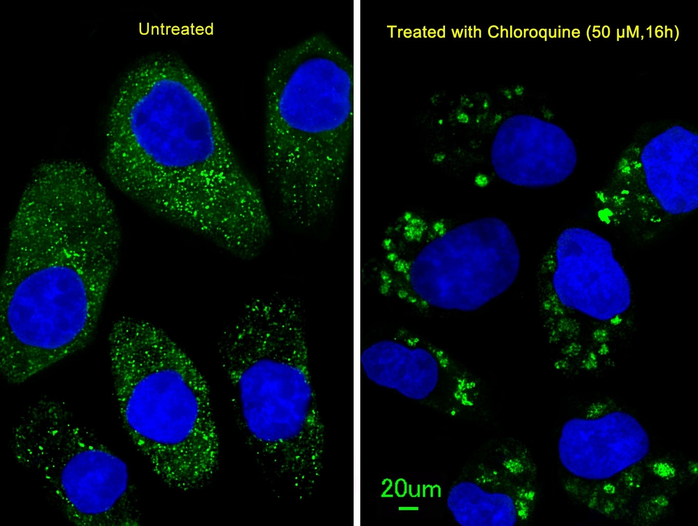
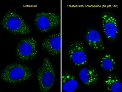
- Immunofluorescence: U251 cells stained with ARG54707 anti-LC3B antibody at 1:25 dilution. U251 cells (right) were treated with Chloroquine (50 µM,16h). DAPI was used to stain the cell nuclear (blue).
- Submitted by
- Arigo (provider)
- Main image
- Experimental details
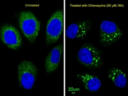
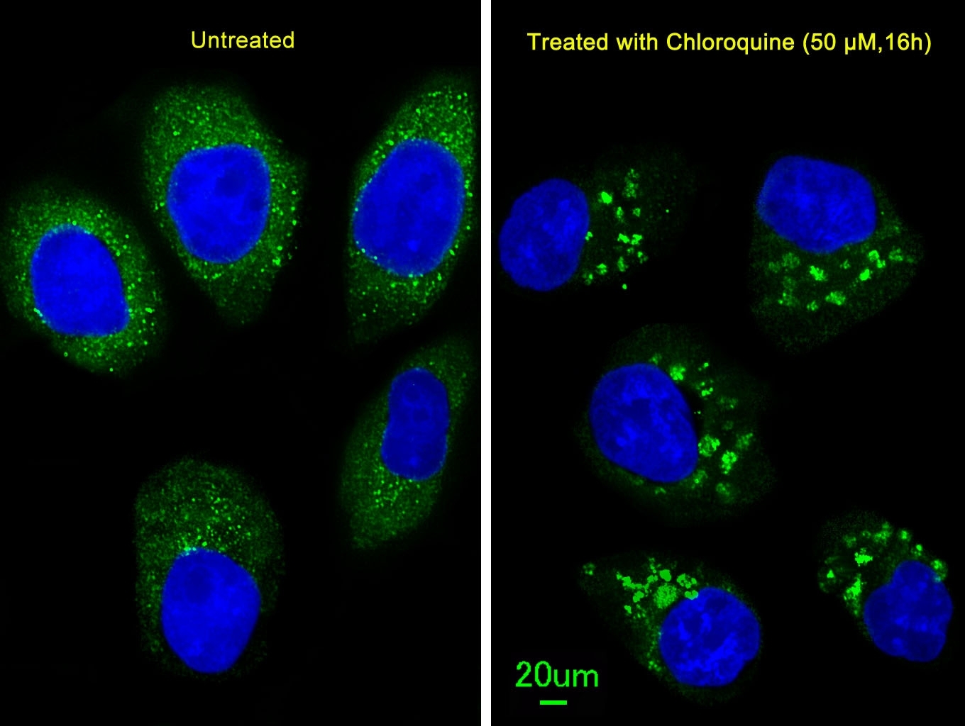
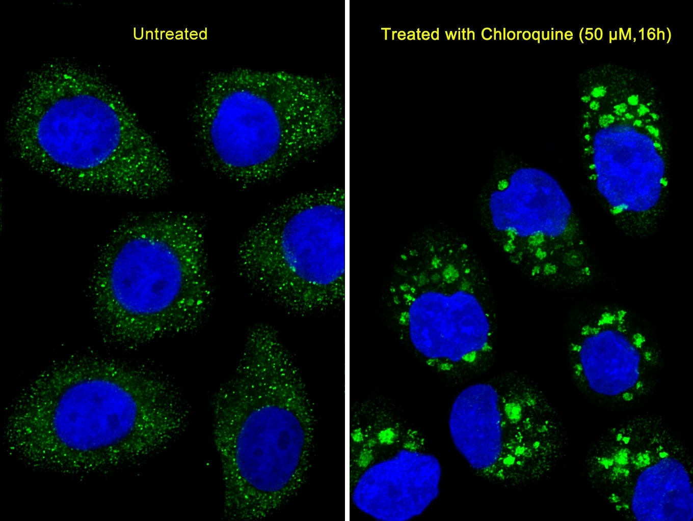
- Immunofluorescence: U251 cells stained with ARG54707 anti-LC3B antibody at 1:100 dilution. U251 cells(right) were treated with Chloroquine (50 µM,16h). DAPI was used to stain the cell nuclear (blue).
- Submitted by
- Arigo (provider)
- Main image
- Experimental details
- Immunofluorescence: U251 cells stained with ARG54707 anti-LC3B antibody at 1:25 dilution. U251 cells(right) were treated with Chloroquine (50 µM,16h). DAPI was used to stain the cell nuclear (blue).
- Submitted by
- Arigo (provider)
- Main image

- Experimental details
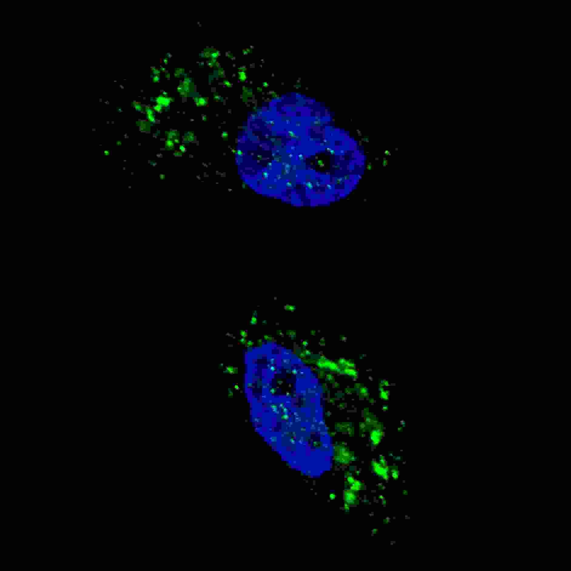
- Immunofluorescence: U251 cells stained with ARG54707 anti-LC3B antibody at 1:100 dilution, 2h, room temperature. U251 cells were treated with Chloroquine (50 µM,16h), then fixed with 4% PFA (20 min), permeabilized with Triton X-100 (0.2%, 30 min). Nuclei were counterstained with Hoechst 33342 (blue) (10 µg/ml, 5 min). LC3 immunoreactivity is localized to autophagic vacuoles in the cytoplasm of U251 cells.
Supportive validation
- Submitted by
- Arigo (provider)
- Main image

- Experimental details
- Immunohistochemistry: Wild-type (Cln3+/+) or homozygous Cln3Äex7/8 (Cln3Äex7/8/Äex7/8) paraffin-embedded brain sections stained with ARG54707 anti-LC3B antibody. Shown are the CA2/CA3 region of hippocampus (Hc) and cerebellum (Cb) from 10-month-old mice. Few immunopositive puncta are present in wild-type sections, whereas homozygous Cln3Äex7/8 sections contain clusters of LC3-positive puncta around pyramidal neurons and Purkinje cells (P). MOL, molecular layer; GCL, granule cell layer. Data courtesy of Dr. Susan Cotman, Massachusets General Hospital.
