Explore
 Validate
 Learn
Learn Western blot
 ELISA
ELISAAntibody data
- Antibody Data
- Antigen structure
- References [0]
- Comments [0]
- Validations
- Western blot [1]
- Immunohistochemistry [1]
- Flow cytometry [1]
Submit
Validation data
Reference
Comment
Report error
- Product number
- 600-401-MN5 - Provider product page

- Provider
- Invitrogen Antibodies
- Product name
- LC3B Polyclonal Antibody
- Antibody type
- Polyclonal
- Antigen
- Synthetic peptide
- Reactivity
- Human
- Host
- Rabbit
- Isotype
- IgG
- Vial size
- 100 µg
- Concentration
- 1 mg/mL
- Storage
- -20° C, Avoid Freeze/Thaw Cycles
No comments: Submit comment
Supportive validation
- Submitted by
- Invitrogen Antibodies (provider)
- Main image
- Experimental details
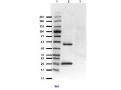
- Western Blot of Rabbit Anti-LC3B Antibody. Lane 1: Opal Prestained Molecular Weight Ladder (p/n Mb-210-0500). Lane 2: MAP1LC3B overexpressing HEK293 (10µg) [+]. Lane 3: HEK293T lysate (10 µg) [-]. Primary Antibody: Anti-LC3B Antibody at 1:1000 overnight at 2-8°C. Secondary Antibody: Goat anti-Rabbit IgG HRP (p/n 611-103-122) at 1:70,000 for 30 minutes. Block: BlockOut Buffer (p/n MB-073). Exposure: 15 sec. Predicted MW: ~14.6, ~63kDa for overexpressing lysates. Observed MW: ~20, 60kDa.

Supportive validation
- Submitted by
- Invitrogen Antibodies (provider)
- Main image
- Experimental details
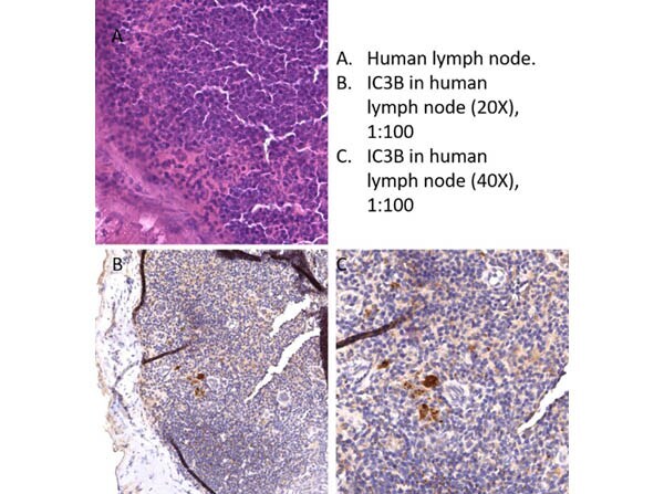
- Immunohistochemistry of Rabbit Anti-LC3B Antibody. Tissue: human lymph node. Fixative: none. Antigen Retrieval: HIER using Cirate Buffer for 20 minutes. Primary Antibody: Anti-LC3B antibody at 1:100 at RT for 30 minutes. Secondary Antibody: Anti-Rabbit Poly-HRP IgG. Ready-to-Use at RT for 8 minutes. Counterstain: Hematoxylin. Substrate: DAB. Results: This antibody showed staining throughout most of the lymph node with focal strong staining. Focal cell staining strongly in the lymph node is the pattern expected.
Supportive validation
- Submitted by
- Invitrogen Antibodies (provider)
- Main image
- Experimental details
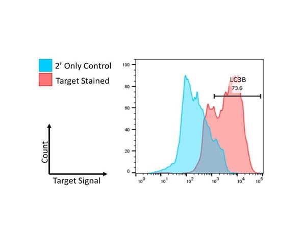
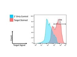
- Flow Cytometry of Rabbit Anti-LC3B Antibody. Cells: U251-MG cells. Primary Antibody: Anti-LC3B at 2.5µg/mL in 100µL FACS buffer for 30 minutes at RT. Secondary Antibody: Donkey anti-Rabbit IgG DyLight™488 (p/n 611-741-127) at 2.5µg/mL in 100µL FACS buffer for 30 minutes at RT. Buffer: FACS/IF buffer (p/n MB-086-0500). Analysis: Distinct positive shifts in fluorescence have been observed in all samples tested following permeabilization of the cell sample. This is indicative of specificity and affinity of each antibody toward its target.