 Explore
 Validate
 Learn
LearnPA5-32254
antibody from Invitrogen Antibodies
Targeting: MAP1LC3B
ATG8F
 Western blot
 Immunocytochemistry Immunoprecipitation Immunohistochemistry Flow cytometry
 Immunoelectron microscopy Other assay
Antibody data
- Antibody Data
- Antigen structure
- References [0]
- Comments [0]
- Validations
- Immunocytochemistry [6]
- Immunoprecipitation [1]
- Immunohistochemistry [2]
- Flow cytometry [2]
- Other assay [3]
Submit
Validation data
Reference
Comment
Report error
- Product number
- PA5-32254 - Provider product page

- Provider
- Invitrogen Antibodies
- Product name
- LC3B Polyclonal Antibody
- Antibody type
- Polyclonal
- Antigen
- Synthetic peptide
- Description
- Recommended positive controls: NT2D1, PC-3, U87-MG, SK-N-SH, mouse brain, Rat brain, HepG2 (untreated), HepG2 (3 µM Thapsigargin treatment for 12 hr), HepG2 (3 µM Thapsigargin treatment for 16hr), HepG2 (3 µM Thapsigargin treatment for 24hr), Huh7 (un-infected), Huh7 (HCV-infected), HeLa, HeLa (50 µM Chloroquine treatment for 24 hr). Predicted reactivity: Zebrafish (100%), Japanese Medaka (100%), Pig (100%), Rhesus Monkey (100%), Chimpanzee (100%), Bovine (100%). Store product as a concentrated solution. Centrifuge briefly prior to opening the vial.
- Reactivity
- Human, Mouse, Rat, Porcine
- Host
- Rabbit
- Isotype
- IgG
- Vial size
- 100 μL
- Concentration
- 0.34 mg/mL
- Storage
- Store at 4°C short term. For long term storage, store at -20°C, avoiding freeze/thaw cycles.
No comments: Submit comment
Supportive validation
- Submitted by
- Invitrogen Antibodies (provider)
- Main image

- Experimental details
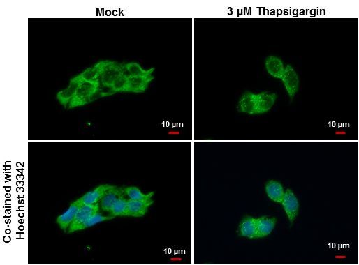
- Immunofluorescent analysis of LC3B1+LC3B2 showing staining in the autophagosome of Hep G2 cells. Hep G2 cells mock (left) and treated with 3µM Thapsigargin for 12 hrs (right) were fixed in ice-cold MeOH for 10 min and permeabilized with ice-cold acetone for 1 min and stained using a LC3B1+LC3B2 polyclonal antibody (Product # PA5-32254) diluted at 1:500. Blue: Hoechst 33342 staining. Scale bar = 10µm.
- Submitted by
- Invitrogen Antibodies (provider)
- Main image

- Experimental details
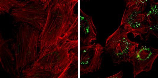
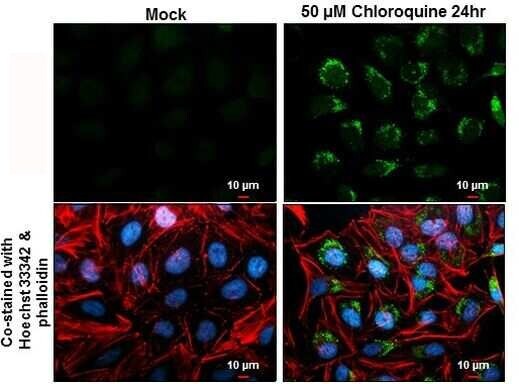
- Immunofluorescent analysis of LC3B1+LC3B2 showing staining in the autophagosome of HeLa cells. HeLa cells mock (left) and treated with 50µM Chloroquine for 24 hr (right) were fixed in 4% paraformaldehyde at RT for 15 min and stained using a LC3B1+LC3B2 polyclonal antibody (Product # PA5-32254) diluted at 1:1000. Red: Phalloidin, a F-actin marker.
- Submitted by
- Invitrogen Antibodies (provider)
- Main image

- Experimental details
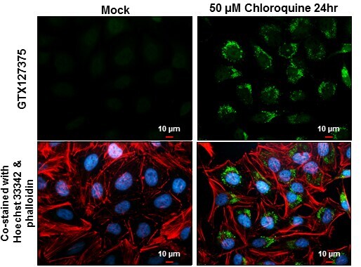
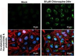
- Immunocytochemistry-Immunofluorescence analysis of LC3B in HeLa cells mock (left) and in HeLa cells treated with 50 µM Chloroquine for 24 hr (right) fixed with 4% paraformaldehyde at RT for 15 min. Green: LC3B Polyclonal Antibody (Product # PA5-32254) diluted at 1:2,000. Red: alpha Tubulin, a cytoskeleton marker, stained by alpha Tubulin antibody. Blue: Hoechst 33342 staining.

- Submitted by
- Invitrogen Antibodies (provider)
- Main image

- Experimental details
- Immunocytochemistry-Immunofluorescence analysis of LC3B was performed in HeLa cells fixed in 4% paraformaldehyde at RT for 15 min. Green: LC3B Polyclonal Antibody (Product # PA5-32254) diluted at 1:200. Red: phalloidin, a cytoskeleton marker. Blue: Hoechst 33342 staining. Scale bar = 10 µm.
- Submitted by
- Invitrogen Antibodies (provider)
- Main image

- Experimental details
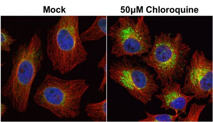
- Immunocytochemistry-Immunofluorescence analysis of LC3B in HeLa cells mock (left) and in HeLa cells treated with 50 µM Chloroquine for 24 hr (right) fixed with 4% paraformaldehyde at RT for 15 min. Green: LC3B Polyclonal Antibody (Product # PA5-32254) diluted at 1:2,000. Red: alpha Tubulin, a cytoskeleton marker, stained by alpha Tubulin antibody. Blue: Hoechst 33342 staining.
- Submitted by
- Invitrogen Antibodies (provider)
- Main image
- Experimental details
- Immunocytochemistry-Immunofluorescence analysis of LC3B was performed in HeLa cells fixed in 4% paraformaldehyde at RT for 15 min. Green: LC3B Polyclonal Antibody (Product # PA5-32254) diluted at 1:200. Red: phalloidin, a cytoskeleton marker. Blue: Hoechst 33342 staining. Scale bar = 10 µm.
Supportive validation
- Submitted by
- Invitrogen Antibodies (provider)
- Main image

- Experimental details
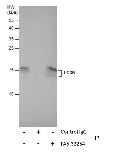
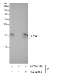
- Immunoprecipitation of LC3B was performed in U87-MG whole cell extracts using 5 µg of LC3B Polyclonal Antibody (Product # PA5-32254). Samples were transferred to a membrane and probed with LC3B Polyclonal Antibody as a primary antibody and an HRP-conjugated anti-Rabbit IgG was used as a secondary antibody.
Supportive validation
- Submitted by
- Invitrogen Antibodies (provider)
- Main image
- Experimental details
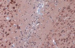
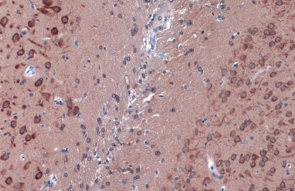
- LC3B Polyclonal Antibody detects LC3B protein at cytoplasm by immunohistochemical analysis. Sample: Paraffin-embedded rat brain. LC3B stained by LC3B Polyclonal Antibody (Product # PA5-32254) diluted at 1:500. Antigen Retrieval: Citrate buffer, pH 6.0, 15 min.
- Submitted by
- Invitrogen Antibodies (provider)
- Main image
- Experimental details
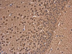
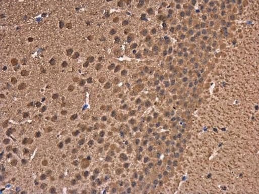
- Immunohistochemistry (Paraffin) analysis of LC3B was performed in paraffin-embedded mouse brain tissue using LC3B Polyclonal Antibody (Product # PA5-32254) at a dilution of 1:500.
Supportive validation
- Submitted by
- Invitrogen Antibodies (provider)
- Main image

- Experimental details
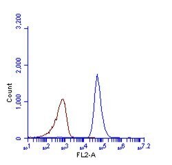
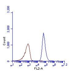
- LC3B Polyclonal Antibody (Product # PA5-32254) detects LC3B protein by flow cytometry analysis. Sample: HeLa cell fixed in 4% paraformaldehyde at 4°C for 5 min. Brown: Unlabelled sample was also used as a control. Blue: LC3B Polyclonal Antibody (Product # PA5-32254) dilution: 1:100. Acquisition of >20,000 events were collected using Argon ion laser (488nm) and 525/30 bandpass filter.

- Submitted by
- Invitrogen Antibodies (provider)
- Main image
- Experimental details
- LC3B Polyclonal Antibody (Product # PA5-32254) detects LC3B protein by flow cytometry analysis. Sample: HeLa cell fixed in 4% paraformaldehyde at 4°C for 5 min. Brown: Unlabelled sample was also used as a control. Blue: LC3B Polyclonal Antibody (Product # PA5-32254) dilution: 1:100. Acquisition of >20,000 events were collected using Argon ion laser (488nm) and 525/30 bandpass filter.
Supportive validation
- Submitted by
- Invitrogen Antibodies (provider)
- Main image
- Experimental details
- NULL
- Submitted by
- Invitrogen Antibodies (provider)
- Main image
- Experimental details
- Immunoprecipitation of LC3B was performed in U87-MG whole cell extracts using 5 µg of LC3B Polyclonal Antibody (Product # PA5-32254). Samples were transferred to a membrane and probed with LC3B Polyclonal Antibody as a primary antibody and an HRP-conjugated anti-Rabbit IgG was used as a secondary antibody.

- Submitted by
- Invitrogen Antibodies (provider)
- Main image
- Experimental details
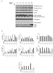
- Figure 3 Low radiation environment switches pKZ1 A11 mouse hybridoma overgrowth cell response from apoptosis toward autophagy. (A) Western blots showing activation of p53 and induction of LC3B-II in LRE (LNGS)-grown pKZ1 A11 mouse hybridoma cells and activation of PARP1 and caspase 3 in RRE (ISS) grown pKZ1 A11 mouse hybridoma cells. (B) Densitometry analysis of western blots shown in (A) .
