 Explore
 Validate
 Learn
Learn Western blot
Antibody data
- Antibody Data
- Antigen structure
- References [1]
- Comments [0]
- Validations
- Western blot [2]
- Immunocytochemistry [2]
- Immunohistochemistry [1]
Submit
Validation data
Reference
Comment
Report error
- Product number
- PA5-35032 - Provider product page

- Provider
- Invitrogen Antibodies
- Product name
- CYP2B6 Polyclonal Antibody
- Antibody type
- Polyclonal
- Antigen
- Synthetic peptide
- Reactivity
- Human
- Host
- Rabbit
- Isotype
- IgG
- Vial size
- 400 μL
- Concentration
- 0.5 mg/mL
- Storage
- Store at 4°C short term. For long term storage, store at -20°C, avoiding freeze/thaw cycles.
Submitted references In vitro toxicological evaluation of NCS-382, a high-affinity antagonist of γ-hydroxybutyrate (GHB) binding.
Vogel KR, Ainslie GR, Roullet JB, McConnell A, Gibson KM
Toxicology in vitro : an international journal published in association with BIBRA 2017 Apr;40:196-202
Toxicology in vitro : an international journal published in association with BIBRA 2017 Apr;40:196-202
No comments: Submit comment
Supportive validation
- Submitted by
- Invitrogen Antibodies (provider)
- Main image
- Experimental details
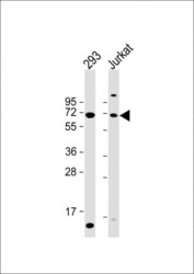
- Western blot analysis of CYP2B6 in various lysates. Samples were incubated with CYP2B6 polyclonal antibody (Product # PA5-35032) using a dilution of 1:2,000 followed by Goat Anti-Rabbit IgG, (H+L), Peroxidase conjugated at a dilution of 1:10,000. Lysates/proteins: 20 µg per lane. Lane 1: 293 whole cell lysate; Lane 2: Jurkat whole cell lysate. Predicted band size: 56 kDa. Blocking/Dilution buffer: 5% NFDM/TBST.
- Submitted by
- Invitrogen Antibodies (provider)
- Main image
- Experimental details
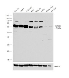
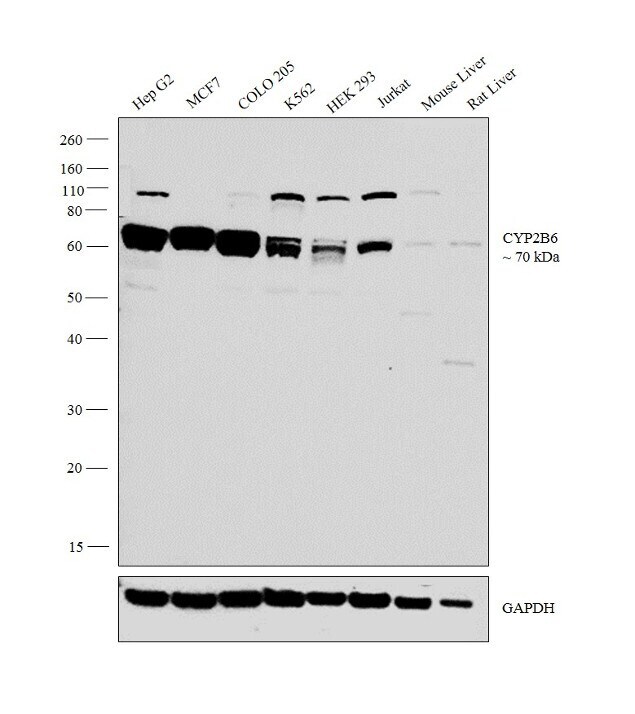
- Western blot analysis was performed on whole cell extracts of Hep G2 (Lane 1), MCF7 (Lane 2), COLO 205 (Lane 3), K562 (Lane 4), HEK 293 (Lane 5), Jurkat (Lane 6), tissue extracts of Mouse Liver (Lane 7) and Rat Liver (Lane 8). The blot was probed with Anti-CYP2B6 Polyclonal Antibody (Product # PA5-35032, 1:1,000 dilution) and detected by chemiluminescence using Goat anti-Rabbit IgG (Heavy Chain) Superclonal™ Secondary Antibody, HRP conjugate (Product # A27036, 0.25 µg/mL, 1:4,000 dilution). A 70 kDa band corresponding to CYP2B6 was detected across the cell lines and tissues tested.
Supportive validation
- Submitted by
- Invitrogen Antibodies (provider)
- Main image

- Experimental details
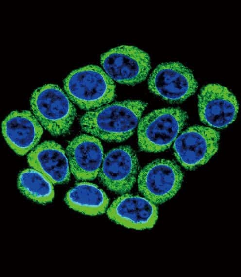
- Immunofluorescent analysis of Cytochrome P450 2B6 in 293 cells using a Cytochrome P450 2B6 polyclonal antibody (Product # PA5-35032) followed by detection using a fluorescent conjugated secondary antibody (green). Nuclei were stained with Dapi (blue).
- Submitted by
- Invitrogen Antibodies (provider)
- Main image
- Experimental details
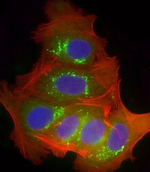
- Immunocytochemistry analysis of CYP2B6 in MCF-7 cells. Samples were incubated with CYP2B6 polyclonal antibody (Product # PA5-35032) using a dilution of 1:25 followed by Dylight® 488-conjugated goat anti-rabbit IgG at a dilution of 1:200 (green). Cells were 4% paraformaldehyde-fixed and 0.1% Triton X-100 permeabilized. Immunofluorescence image showing endosomes staining on MCF-7 cell line. Cytoplasmic actin is detected with Dylight® 554 Phalloidin at 1:100 dilution (red). The nuclear counter stain is DAPI (blue).

Supportive validation
- Submitted by
- Invitrogen Antibodies (provider)
- Main image

- Experimental details
- Immunohistochemistry analysis of CYP2B6 in formalin fixed and paraffin embedded human hepatocarcinoma. Samples were incubated with CYP2B6 polyclonal antibody (Product # PA5-35032) followed by peroxidase conjugation of the secondary antibody and DAB staining. This data demonstrates the use of this antibody for immunohistochemistry. Clinical relevance has not been evaluated.
