Explore
 Validate
 Learn
Learn Western blot
 Immunocytochemistry
Antibody data
- Antibody Data
- Antigen structure
- References [1]
- Comments [0]
- Validations
- Western blot [2]
- Immunohistochemistry [11]
Submit
Validation data
Reference
Comment
Report error
- Product number
- NBP1-89517 - Provider product page

- Provider
- Novus Biologicals
- Proper citation
- Novus Cat#NBP1-89517, RRID:AB_11033728
- Product name
- Rabbit Polyclonal NUDC Antibody
- Antibody type
- Polyclonal
- Description
- Immunogen affinity purified. Specificity of human, mouse, rat NUDC antibody verified on a Protein Array containing target protein plus 383 other non-specific proteins.
- Reactivity
- Human, Mouse, Rat
- Host
- Rabbit
- Isotype
- IgG
- Vial size
- 0.1 ml
- Storage
- Store at 4C short term. Aliquot and store at -20C long term. Avoid freeze-thaw cycles.
Submitted references Immunofluorescence and fluorescent-protein tagging show high correlation for protein localization in mammalian cells.
Stadler C, Rexhepaj E, Singan VR, Murphy RF, Pepperkok R, Uhlén M, Simpson JC, Lundberg E
Nature methods 2013 Apr;10(4):315-23
Nature methods 2013 Apr;10(4):315-23
No comments: Submit comment
Supportive validation
- Submitted by
- Novus Biologicals (provider)
- Main image
- Experimental details
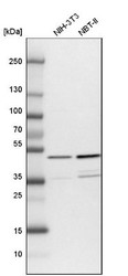
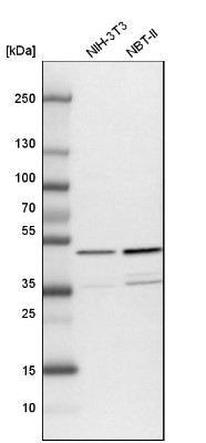
- Western Blot: NUDC Antibody [NBP1-89517] - Analysis in mouse cell line NIH-3T3 and rat cell line NBT-II.
- Submitted by
- Novus Biologicals (provider)
- Main image
- Experimental details
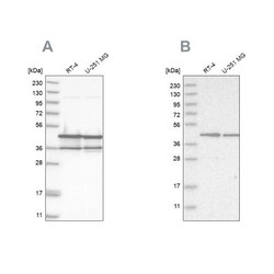
- Western Blot: NUDC Antibody [NBP1-89517] - Analysis using Anti-NUDC antibody NBP1-89517 (A) shows similar pattern to independent antibody NBP1-89510 (B).

Supportive validation
- Submitted by
- Novus Biologicals (provider)
- Main image
- Experimental details
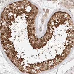
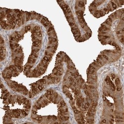
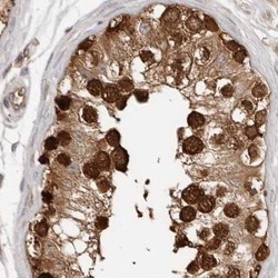
- Immunohistochemistry-Paraffin: NUDC Antibody [NBP1-89517] - Staining of human testis shows distinct nuclear and cytoplasmic positivity in seminiferus duct cells.

- Submitted by
- Novus Biologicals (provider)
- Main image
- Experimental details
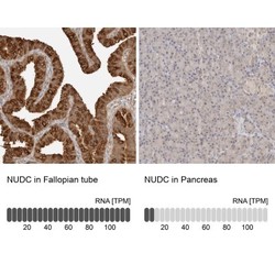
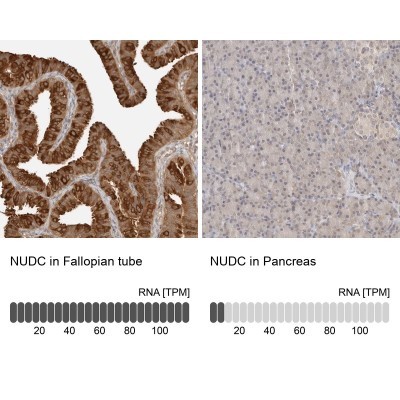
- Immunohistochemistry-Paraffin: NUDC Antibody [NBP1-89517] - Staining of human fallopian tube shows high expression.

- Submitted by
- Novus Biologicals (provider)
- Main image
- Experimental details
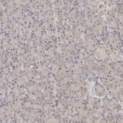
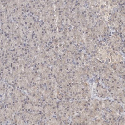
- Immunohistochemistry-Paraffin: NUDC Antibody [NBP1-89517] - Staining of human pancreas shows low expression as expected.
- Submitted by
- Novus Biologicals (provider)
- Main image
- Experimental details
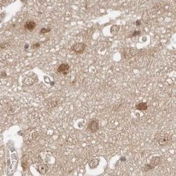
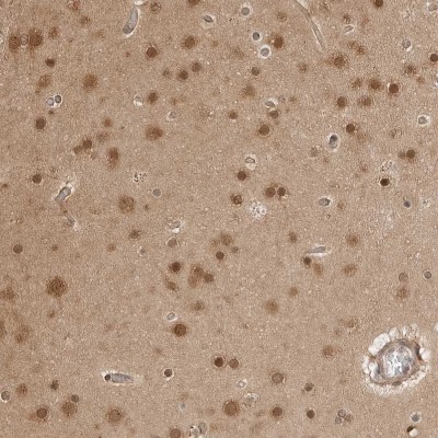
- Immunohistochemistry-Paraffin: NUDC Antibody [NBP1-89517] - Staining of human cerebral cortex.

- Submitted by
- Novus Biologicals (provider)
- Main image
- Experimental details
- Immunohistochemistry-Paraffin: NUDC Antibody [NBP1-89517] - Staining in human fallopian tube and pancreas tissues using anti-NUDC antibody. Corresponding NUDC RNA-seq data are presented for the same tissues.
- Submitted by
- Novus Biologicals (provider)
- Main image
- Experimental details
- Immunohistochemistry-Paraffin: NUDC Antibody [NBP1-89517] - Staining of human endometrium.

- Submitted by
- Novus Biologicals (provider)
- Main image
- Experimental details
- Immunohistochemistry-Paraffin: NUDC Antibody [NBP1-89517] - Staining of human testis.

- Submitted by
- Novus Biologicals (provider)
- Main image
- Experimental details
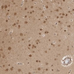
- Immunohistochemistry-Paraffin: NUDC Antibody [NBP1-89517] - Staining of human cerebral cortex shows weak to moderate cytoplasmic and nuclear positivity in neurons.
- Submitted by
- Novus Biologicals (provider)
- Main image
- Experimental details
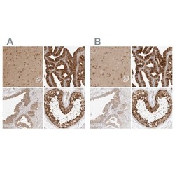
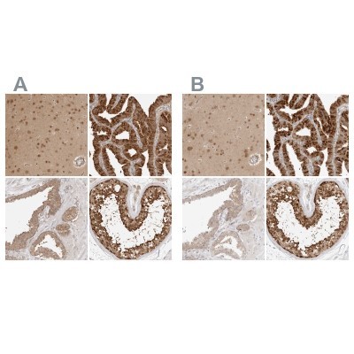
- Immunohistochemistry-Paraffin: NUDC Antibody [NBP1-89517] - Staining of human cerebral cortex, fallopian tube, prostate and testis using Anti-NUDC antibody NBP1-89517 (A) shows similar protein distribution across tissues to independent antibody NBP1-89510 (B).
- Submitted by
- Novus Biologicals (provider)
- Main image

- Experimental details
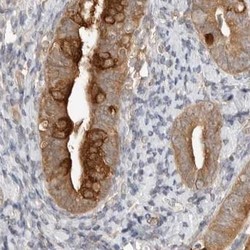
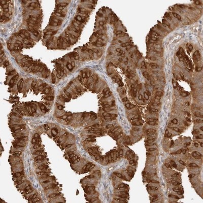
- Immunohistochemistry-Paraffin: NUDC Antibody [NBP1-89517] - Staining of human Fallopian tube shows strong cytoplasmic positivity in glandular cells.
- Submitted by
- Novus Biologicals (provider)
- Main image

- Experimental details
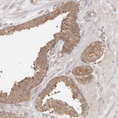
- Immunohistochemistry-Paraffin: NUDC Antibody [NBP1-89517] - Staining of human prostate shows moderate cytoplasmic positivity in glandular cells.