Explore
 Validate
 Learn
Learn Western blot
Antibody data
- Antibody Data
- Antigen structure
- References [0]
- Comments [0]
- Validations
- Western blot [4]
- Immunocytochemistry [2]
- Immunohistochemistry [4]
Submit
Validation data
Reference
Comment
Report error
- Product number
- PA5-53317 - Provider product page

- Provider
- Invitrogen Antibodies
- Product name
- HADHA Polyclonal Antibody
- Antibody type
- Polyclonal
- Antigen
- Recombinant full-length protein
- Description
- Immunogen sequence: IEYLEEVAIT FAKGLADKKI SPKRDKGLVE KLTAYAMTIP FVRQQVYKKV EEKVRKQTKG LYPAPLKIID VVKTGIEQGS DAGYLCESQK FGELVMTKES KALMGLYHGQ VLCKKNKFGA PQKDVKHLAI LGAGLMGAGI AQVSVDK
- Concentration
- 0.1 mg/mL
No comments: Submit comment
Supportive validation
- Submitted by
- Invitrogen Antibodies (provider)
- Main image
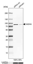
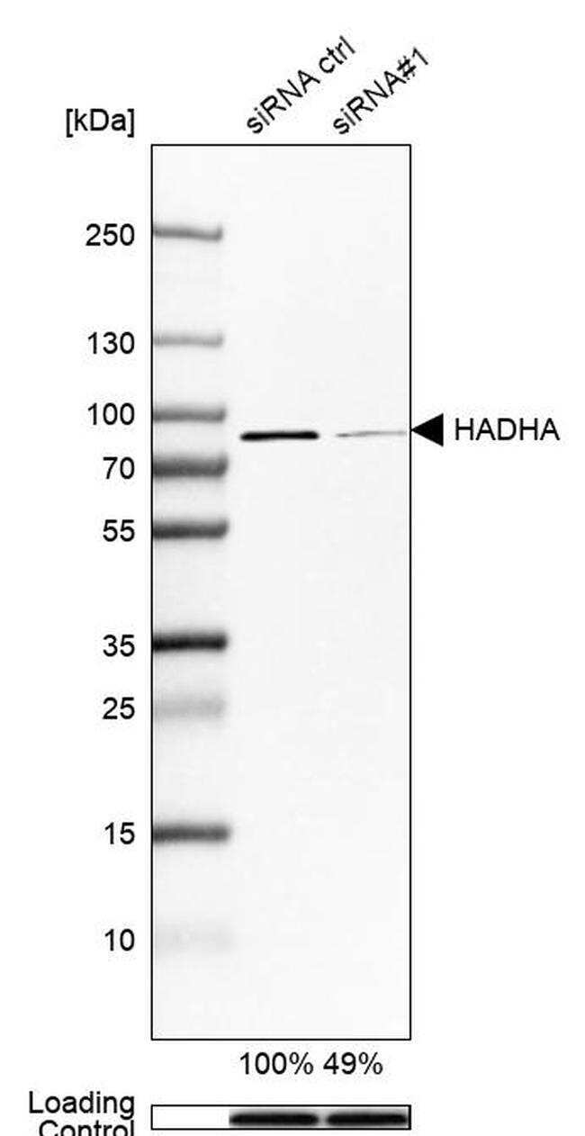
- Experimental details
- Western blot analysis of HADHA in Hep-G2 cells transfected with control siRNA, target specific siRNA probe #1, using a HADHA Polyclonal Antibody (Product # PA5-53317). Remaining relative intensity is presented. Loading control: Anti-GAPDH.
- Submitted by
- Invitrogen Antibodies (provider)
- Main image

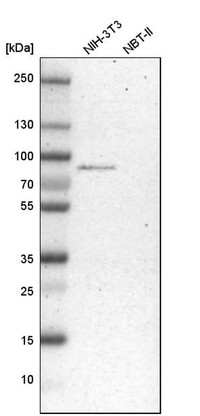
- Experimental details
- Western blot analysis of HADHA in mouse cell line NIH-3T3 and rat cell line NBT-II using a HADHA Polyclonal Antibody (Product # PA5-53317).
- Submitted by
- Invitrogen Antibodies (provider)
- Main image
- Experimental details
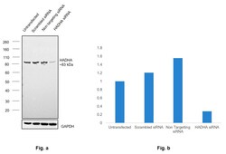
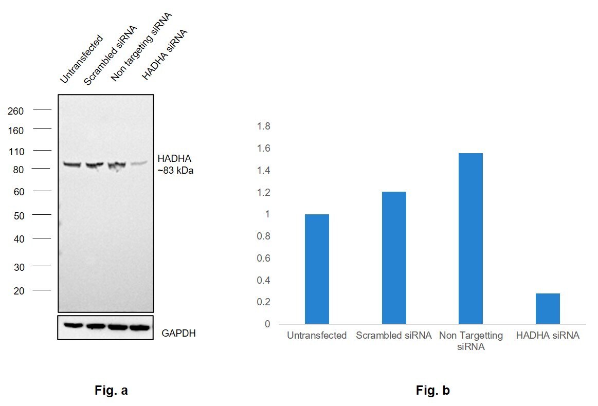
- Knockdown of HADHA was achieved by transfecting Hep G2 with HADHA specific siRNAs (Silencer® select Product # s6439, s6440). Western Blot analysis (Fig. a) was performed using Whole cell extracts from the HADHA knockdown cells (lane 3), non-targeting scrambled siRNA transfected cells (lane 2) and untransfected cells (lane 1). The blot was probed with HADHA Polyclonal Antibody (Product # PA5-53317, 0.04 µg/mL ) and Goat anti-Rabbit IgG (H+L) Superclonal™ Recombinant Secondary Antibody, HRP (Product # A27036, 1:4000). Densitometric analysis of this Western Blot is shown in histogram (Fig. b). Decrease in signal upon siRNA mediated knock down confirms that antibody is specific to HADHA. Non targeting siRNA for HepG2 was also run and quantified as shown.
- Submitted by
- Invitrogen Antibodies (provider)
- Main image
- Experimental details
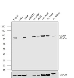
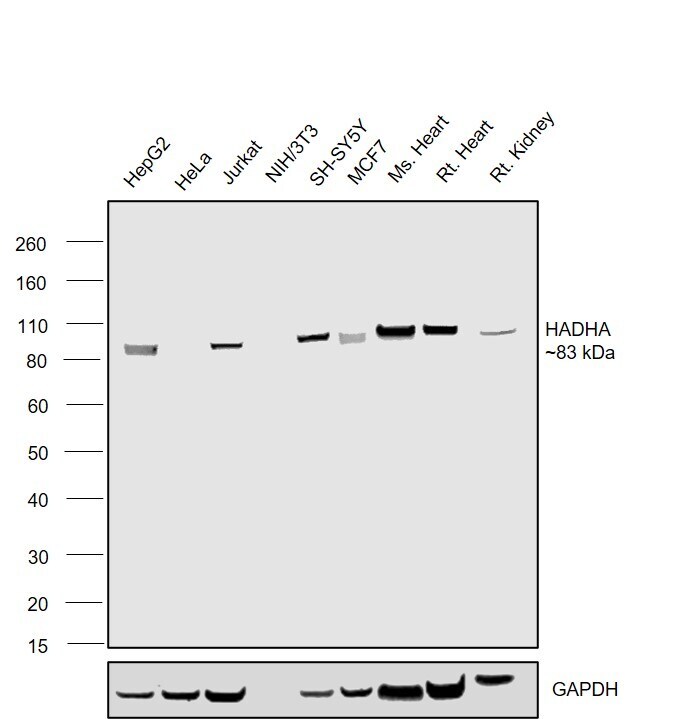
- Western Blot was performed using Anti-HADHA Polyclonal Antibody (Product # PA5-53317) and a 83 kDa band corresponding to HADHA was observed across cell lines and tissues tested . Whole cell extracts (30 µg lysate) of Hep G2 (Lane 1), HeLa (Lane 2), Jurkat (Lane 3), NIH/3T3 (Lane 4), SH-SY5Y (Lane 5), MCF7 (Lane 6), Mouse Heart (Lane 7), Rat Heart (Lane 8), Rat Kidney (Lane 9) were electrophoresed using NuPAGE™ 4-12% Bis-Tris Protein Gel (Product # NP0322BOX). Resolved proteins were then transferred onto a nitrocellulose membrane (Product # LC2001) by iBlot® 2 Dry Blotting System (Product # IB21001). The blot was probed with the primary antibody (0.04 µg/mL) and detected by chemiluminescence with Goat anti-Rabbit IgG (H+L) Superclonal™ Recombinant Secondary Antibody, HRP (Product # A27036, 1:4000) using the iBright FL 1000 (Product # A32752). Chemiluminescent detection was performed using Novex® ECL Chemiluminescent Substrate Reagent Kit (Product # WP20005).
Supportive validation
- Submitted by
- Invitrogen Antibodies (provider)
- Main image

- Experimental details
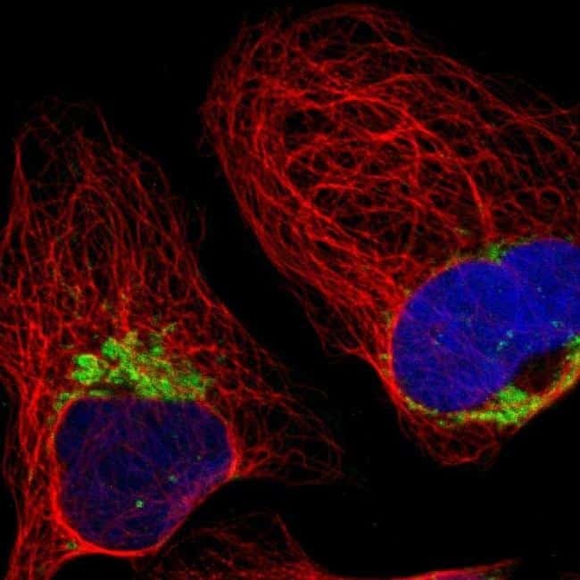
- Immunofluorescent staining of HADHA in human cell line U-2 OS shows positivity in mitochondria. Samples were probed using a HADHA Polyclonal Antibody (Product # PA5-53317).
- Submitted by
- Invitrogen Antibodies (provider)
- Main image
- Experimental details
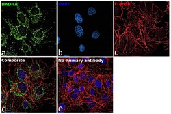
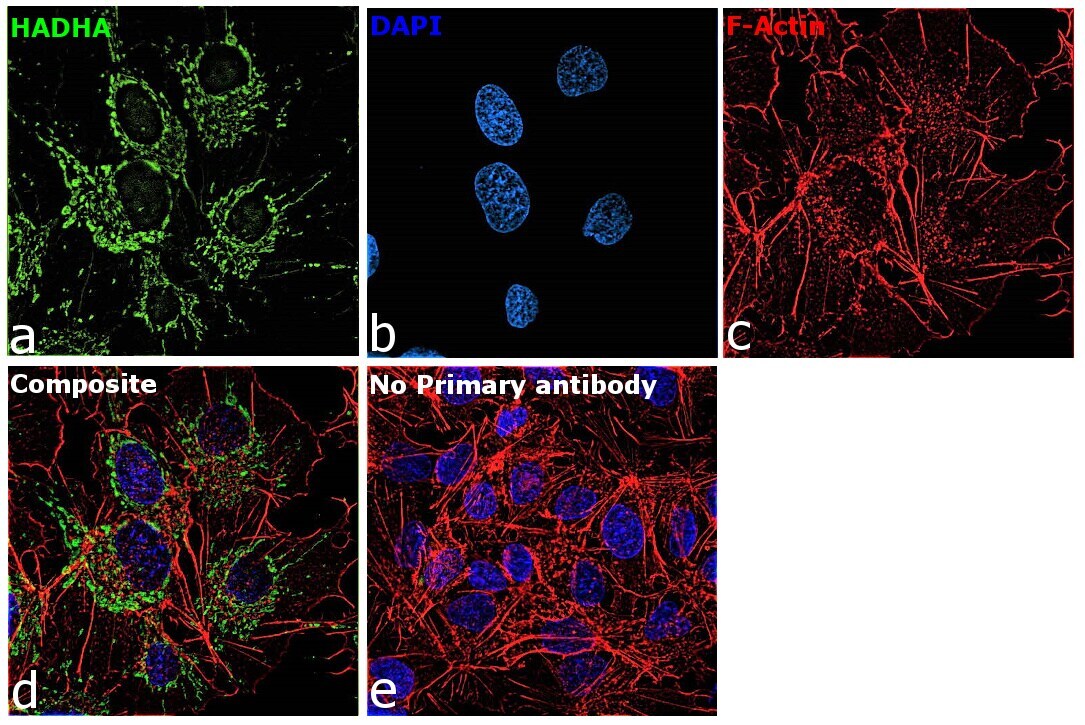
- Immunofluorescence analysis of HADHA was performed using 70% confluent log phase Hep G2 cells. The cells were fixed with 4% paraformaldehyde for 10 minutes, permeabilized with 0.1% Triton™ X-100 for 15 minutes, and blocked with 2% BSA for 45 minutes at room temperature. The cells were labeled with HADHA Polyclonal Antibody (Product # PA5-53317) at 1:100 dilution in 0.1% BSA, incubated at 4 degree celsius overnight and then labeled with Donkey anti-Rabbit IgG (H+L) Highly Cross-Adsorbed Secondary Antibody, Alexa Fluor Plus 488 (Product # A32790), (1:2000), for 45 minutes at room temperature (Panel a: Green). Nuclei (Panel b:Blue) were stained with ProLong™ Diamond Antifade Mountant with DAPI (Product # P36962). F-actin (Panel c: Red) was stained with Rhodamine Phalloidin (Product # R415, 1:300). Panel d represents the merged image showing mitochondrial localization. Panel e represents control cells with no primary antibody to assess background. The images were captured at 60X magnification.
Supportive validation
- Submitted by
- Invitrogen Antibodies (provider)
- Main image
- Experimental details
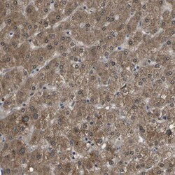
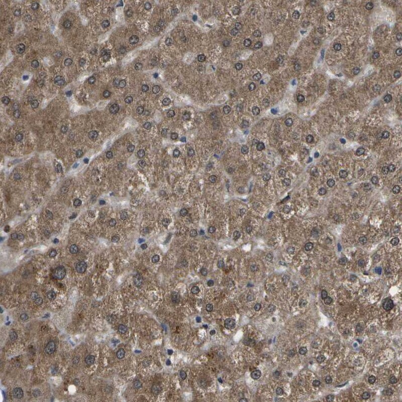
- Immunohistochemical staining of HADHA in human liver using HADHA Polyclonal Antibody (Product # PA5-53317) shows strong cytoplasmic positivity in hepatocytes.
- Submitted by
- Invitrogen Antibodies (provider)
- Main image
- Experimental details
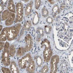
- Immunohistochemical staining of HADHA in human kidney using HADHA Polyclonal Antibody (Product # PA5-53317) shows moderate to strong cytoplasmic positivity in cells in tubules.

- Submitted by
- Invitrogen Antibodies (provider)
- Main image
- Experimental details
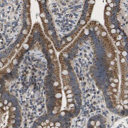
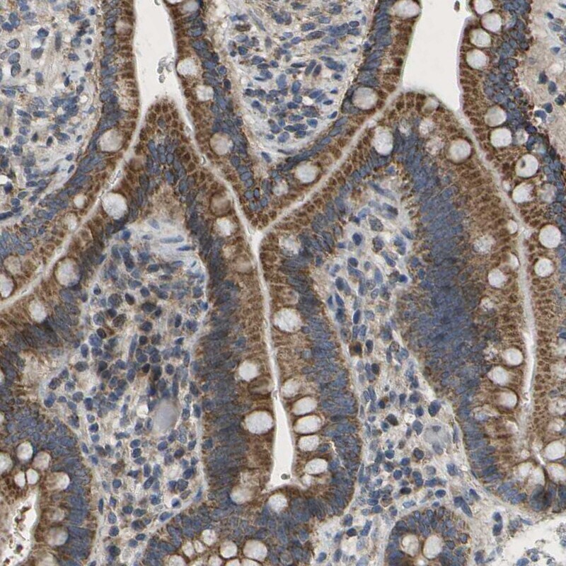
- Immunohistochemical staining of HADHA in human small intestine using HADHA Polyclonal Antibody (Product # PA5-53317) shows strong granular cytoplasmic positivity in glandular cells.
- Submitted by
- Invitrogen Antibodies (provider)
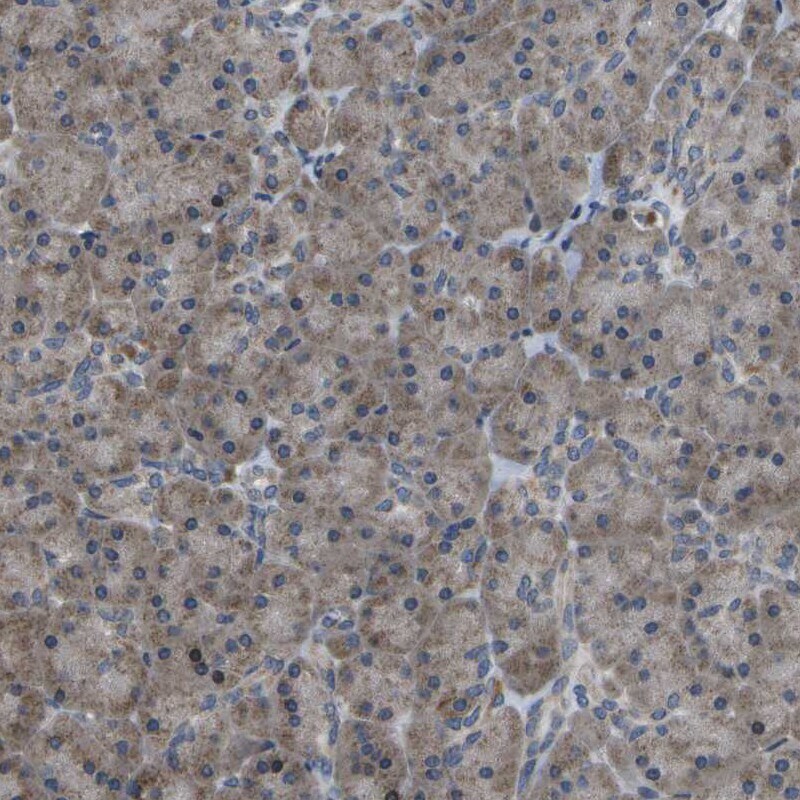
- Main image
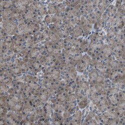
- Experimental details
- Immunohistochemical staining of HADHA in human pancreas using HADHA Polyclonal Antibody (Product # PA5-53317) shows moderate cytoplasmic positivity in exocrine glandular cells.