Explore
 Validate
 Learn
Learn Western blot
 Immunocytochemistry
 Immunohistochemistry
Antibody data
- Antibody Data
- Antigen structure
- References [1]
- Comments [0]
- Validations
- Western blot [1]
- Immunocytochemistry [1]
Submit
Validation data
Reference
Comment
Report error
- Product number
- HPA015536 - Provider product page

- Provider
- Atlas Antibodies
- Proper citation
- Atlas Antibodies Cat#HPA015536, RRID:AB_1850473
- Product name
- Anti-HADHA
- Antibody type
- Polyclonal
- Description
- Polyclonal Antibody against Human HADHA, Gene description: hydroxyacyl-CoA dehydrogenase/3-ketoacyl-CoA thiolase/enoyl-CoA hydratase (trifunctional protein), alpha subunit, Alternative Gene Names: GBP, LCEH, LCHAD, MTPA, Validated applications: WB, IHC, ICC, Uniprot ID: P40939, Storage: Store at +4°C for short term storage. Long time storage is recommended at -20°C.
- Reactivity
- Human, Mouse
- Host
- Rabbit
- Conjugate
- Unconjugated
- Isotype
- IgG
- Vial size
- 100 µl
- Concentration
- 0.1 mg/ml
- Storage
- Store at +4°C for short term storage. Long time storage is recommended at -20°C.
- Handling
- The antibody solution should be gently mixed before use.
Submitted references Expression of miR-33 from an SREBP2 Intron Inhibits Cholesterol Export and Fatty Acid Oxidation*
Gerin I, Clerbaux L, Haumont O, Lanthier N, Das A, Burant C, Leclercq I, MacDougald O, Bommer G
Journal of Biological Chemistry 2010;285(44):33652-33661
Journal of Biological Chemistry 2010;285(44):33652-33661
No comments: Submit comment
Enhanced validation
- Submitted by
- Atlas Antibodies (provider)
- Enhanced method
- Genetic validation
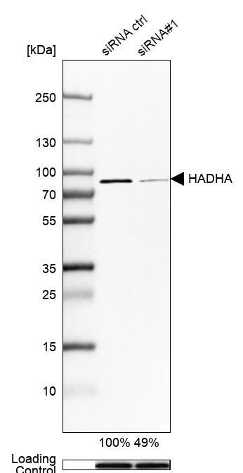
- Main image

- Experimental details
- Western blot analysis in Hep-G2 cells transfected with control siRNA, target specific siRNA probe #1, using Anti-HADHA antibody. Remaining relative intensity is presented. Loading control: Anti-GAPDH.
- Sample type
- Human
- Protocol
- Protocol
Supportive validation
- Submitted by
- Atlas Antibodies (provider)
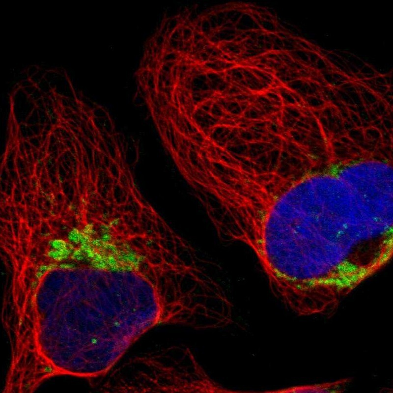
- Main image

- Experimental details
- Immunofluorescent staining of human cell line U-2 OS shows localization to mitochondria.
- Sample type
- Human