Explore
 Validate
 Learn
Learn Western blot
Antibody data
- Antibody Data
- Antigen structure
- References [0]
- Comments [0]
- Validations
- Western blot [2]
- Immunohistochemistry [3]
- Flow cytometry [1]
Submit
Validation data
Reference
Comment
Report error
- Product number
- NBP2-29954 - Provider product page

- Provider
- Novus Biologicals
- Product name
- Rabbit Polyclonal PLA2G7/PAF-AH/Lp-PLA2 Antibody
- Antibody type
- Polyclonal
- Description
- Protein A purified.
- Reactivity
- Human
- Host
- Rabbit
- Isotype
- IgG
- Vial size
- 0.4 ml
- Concentration
- 1.3 mg/ml
- Storage
- Store at 4C short term. Aliquot and store at -20C long term. Avoid freeze-thaw cycles.
No comments: Submit comment
Supportive validation
- Submitted by
- Novus Biologicals (provider)
- Main image

- Experimental details
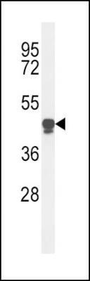
- Western Blot: PLA2G7/PAF-AH/Lp-PLA2 Antibody [NBP2-29954] - Western blot analysis of PLA2G7 Antibody (Center) (NBP2-29954) in HL-60 cell line lysates (35ug/lane). PLA2G7 (arrow) was detected using the purified Pab.
- Submitted by
- Novus Biologicals (provider)
- Main image
- Experimental details
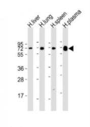
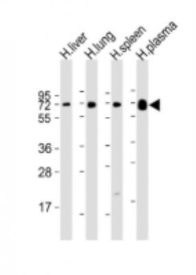
- Western Blot: PLA2G7/PAF-AH/Lp-PLA2 Antibody [NBP2-29954] - Anti-PLA2G7 Antibody (Center) at 1:2000 dilution Lane 1: human liver lysate Lane 2: human lung lysate Lane 3: human spleen lysate Lane 4: human plasma lysate Lysates/proteins at 20 ug per lane. Secondary Goat Anti-Rabbit IgG, (H+L), Peroxidase conjugated at 1/10000 dilution. Predicted band size : 50 kDa Blocking/Dilution buffer: 5% NFDM/TBST.
Supportive validation
- Submitted by
- Novus Biologicals (provider)
- Main image

- Experimental details
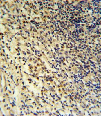
- Immunohistochemistry: PLA2G7/PAF-AH/Lp-PLA2 Antibody [NBP2-29954] - IHC analysis in formalin fixed and paraffin embedded tonsil followed by peroxidase conjugation of the secondary antibody and DAB staining. This data demonstrates the use of the PLA2G7 Antibody (Center) for immunohistochemistry. Clinical relevance has not been evaluated.
- Submitted by
- Novus Biologicals (provider)
- Main image
- Experimental details
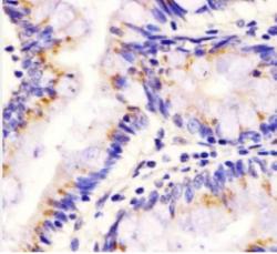
- Immunohistochemistry-Paraffin: PLA2G7/PAF-AH/Lp-PLA2 Antibody [NBP2-29954] - Staining PLA2G7 in human colon tissue sections by Immunohistochemistry (IHC-P - paraformaldehyde-fixed, paraffin-embedded sections). Tissue was fixed with formaldehyde and blocked with 3% BSA for 0. 5 hour at room temperature; antigen retrieval was by heat mediation with a citrate buffer (pH6). Samples were incubated with primary antibody (1/25) for 1 hours at 37 degrees C. A undiluted biotinylated goat polyvalent antibody was used as the secondary antibody.

- Submitted by
- Novus Biologicals (provider)
- Main image
- Experimental details
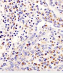
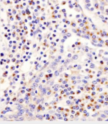
- Immunohistochemistry-Paraffin: PLA2G7/PAF-AH/Lp-PLA2 Antibody [NBP2-29954] - Staining PLA2G7 in human tonsil tissue sections by Immunohistochemistry (IHC-P - paraformaldehyde-fixed, paraffin-embedded sections). Tissue was fixed with formaldehyde and blocked with 3% BSA for 0. 5 hour at room temperature; antigen retrieval was by heat mediation with a citrate buffer (pH6). Samples were incubated with primary antibody (1/25) for 1 hours at 37 degrees C. A undiluted biotinylated goat polyvalent antibody was used as the secondary antibody.
Supportive validation
- Submitted by
- Novus Biologicals (provider)
- Main image
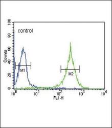
- Experimental details
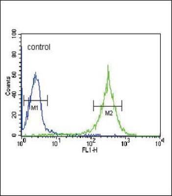
- Flow Cytometry: PLA2G7/PAF-AH/Lp-PLA2 Antibody [NBP2-29954] - Flow cytometric analysis of HL-60 cells (right histogram) compared to a negative control cell (left histogram).FITC-conjugated goat-anti-rabbit secondary antibodies were used for the analysis.