Explore
 Validate
 Learn
Learn Western blot
 ELISA
ELISA Immunocytochemistry
Antibody data
- Antibody Data
- Antigen structure
- References [0]
- Comments [0]
- Validations
- Immunocytochemistry [1]
- Flow cytometry [1]
Submit
Validation data
Reference
Comment
Report error
- Product number
- MA5-51710 - Provider product page

- Provider
- Invitrogen Antibodies
- Product name
- MAGEA3 Chimeric Recombinant Rabbit Monoclonal Antibody (21B4)
- Antibody type
- Monoclonal
- Antigen
- Other
- Description
- This antibody binds human MAGEA3 and MAGEA6, but does not cross react with other members of the MAGE family like MAGE-A1, MAGE-A4 and MAGE-A10. MAGE-A3 is a tumor specific antigen and it is expressed on many tumors like melanoma, non-small cell lung cancer, hematologic malignancies, and others.
- Reactivity
- Human
- Host
- Rabbit
- Isotype
- IgG
- Antibody clone number
- 21B4
- Vial size
- 200 μg
- Concentration
- 1 mg/mL
- Storage
- Store at 4°C short term. For long term storage, store at -20°C, avoiding freeze/thaw cycles.
No comments: Submit comment
Supportive validation
- Submitted by
- Invitrogen Antibodies (provider)
- Main image
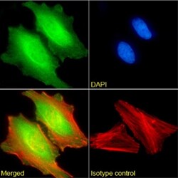
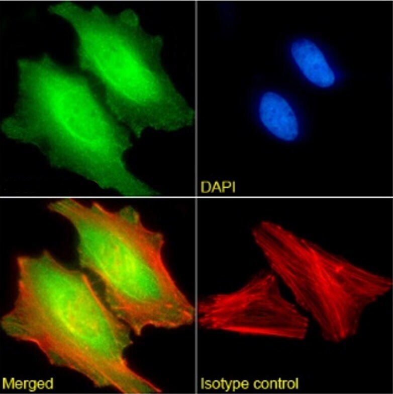
- Experimental details
- Immunocytochemistry analysis of MAGEA3 in paraformaldehyde fixed HeLa cells using MAGEA3 Chimeric Recombinant Rabbit Monoclocal Antibody (Product # MA5-51710) at a 1:1,000 dilution for 1h followed by Alexa Fluor 488 secondary antibody (1:1,500 dilution), showing nuclear staining. The nuclear stain is DAPI (blue). Panels show from left-right, top-bottom: primary antibody, DAPI, merged channels and an isotype control. The isotype control was anti-fluorescein antibody (Product # MA5-47752) followed by staining with Alexa Fluor 488 secondary antibody.
Supportive validation
- Submitted by
- Invitrogen Antibodies (provider)
- Main image
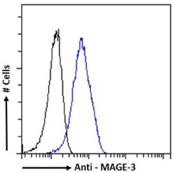
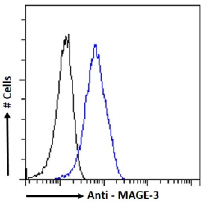
- Experimental details
- Flow cytometry analysis of MAGEA3 in HeLa cells using MAGEA3 Chimeric Recombinant Rabbit Monoclocal Antibody (Product # MA5-51710). Samples were stained with an isotype control (Product # MA5-47752, black line) or primary antibody (Product # MA5-51710, blue line) at a concentration of 1 µg/mL for 30 mins at RT. After washing, bound antibody was detected using Alexa Fluor 488 conjugated goat anti-rabbit IgG and cells analysed on a flow-cytometer.