 Explore
 Validate
 Learn
Learn Western blot
Antibody data
- Antibody Data
- Antigen structure
- References [0]
- Comments [0]
- Validations
- Western blot [5]
- Immunohistochemistry [5]
Submit
Validation data
Reference
Comment
Report error
- Product number
- PA5-57019 - Provider product page

- Provider
- Invitrogen Antibodies
- Product name
- UGP2 Polyclonal Antibody
- Antibody type
- Polyclonal
- Antigen
- Recombinant full-length protein
- Description
- Immunogen sequence: TVPLVKLGSS FTKVQDYLRR FESIPDMLEL DHLTVSGDVT FGKNVSLKGT VIIIANHGDR IDIPPGAVLE NKIVSGNL
- Concentration
- 0.05 mg/mL
No comments: Submit comment
Supportive validation
- Submitted by
- Invitrogen Antibodies (provider)
- Main image

- Experimental details
- Western blot analysis of UGP2 in Lane 1: Mouse liver tissue. lysate; Lane 2: Rat liver tissue. lysate. Samples were probed using an UGP2 Polyclonal Antibody (Product # PA5-57019).

- Submitted by
- Invitrogen Antibodies (provider)
- Main image
- Experimental details
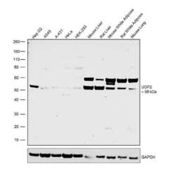
- Western blot was performed using Anti-UGP2 Polyclonal Antibody (Product # PA5-57019) and a 56 kDa band corresponding to UGP2 was observed across all the cell lines and tissues tested. Whole cell extracts (30 µg lysate) of Hep G2 (Lane 1), A549 (Lane 2), A-431 (Lane 3), HeLa (Lane 4), HEK-293 (Lane 5), tissue extracts of Mouse Liver (Lane 6), Rat Liver (Lane 7), Mouse White Adipose (Lane 8), Rat White Adipose (Lane 9) and Mouse Lung (Lane 10) were electrophoresed using NuPAGE™ 4-12% Bis-Tris Protein Gel (Product # NP0322BOX). Resolved proteins were then transferred onto a nitrocellulose membrane (Product # IB23001) by iBlot® 2 Dry Blotting System (Product # IB21001). The blot was probed with the primary antibody (0.4 ug/ml) and detected by chemiluminescence using Goat Anti-Rabbit IgG Secondary Antibody, HRP conjugate (Product # A27036, 1:4000 dilution) using the iBright FL 1000 (Product # A32752). Chemiluminescent detection was performed using Novex® ECL Chemiluminescent Substrate Reagent Kit (Product # WP20005).

- Submitted by
- Invitrogen Antibodies (provider)
- Main image
- Experimental details
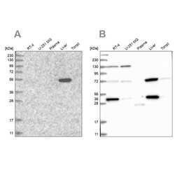
- Western blot analysis of UGP2 using UGP2 Polyclonal Antibody (Product # PA5-57019) (A) shows similar pattern to an independent UGP2 Polyclonal Antibody (B).

- Submitted by
- Invitrogen Antibodies (provider)
- Main image
- Experimental details
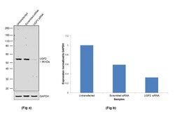
- Knockdown of UGP2 was achieved by transfecting Hep G2 cells with UGP2 specific siRNA (Silencer® select Product # S14645). Western blot analysis (Fig. a) was performed using whole cell extracts from the UGP2 knockdown cells (Lane 3), non-specific scrambled siRNA transfected cells (Lane 2) and untransfected cells (Lane 1). The blot was probed with UGP2 Polyclonal Antibody (Product # PA5-57019, 0.4 ug/ml) and Goat anti-Rabbit IgG (H+L) Superclonal™ Secondary Antibody, HRP conjugate (Product # A27036, 1:4000 dilution). Densitometric analysis of this western blot is shown in histogram (Fig. b). Loss of signal upon siRNA mediated knock down confirms that antibody is specific to UGP2.

- Submitted by
- Invitrogen Antibodies (provider)
- Main image

- Experimental details
- Western blot was performed using Anti-UGP2 Polyclonal Antibody (Product # PA5-57019) and a 56 kDa band corresponding to UGP2 was observed across all the cell lines and tissues tested. Whole cell extracts (30 µg lysate) of Hep G2 (Lane 1), A549 (Lane 2), A-431 (Lane 3), HeLa (Lane 4), HEK-293 (Lane 5), tissue extracts of Mouse Liver (Lane 6), Rat Liver (Lane 7), Mouse White Adipose (Lane 8), Rat White Adipose (Lane 9) and Mouse Lung (Lane 10) were electrophoresed using NuPAGE™ 4-12% Bis-Tris Protein Gel (Product # NP0322BOX). Resolved proteins were then transferred onto a nitrocellulose membrane (Product # IB23001) by iBlot® 2 Dry Blotting System (Product # IB21001). The blot was probed with the primary antibody (0.4 ug/ml) and detected by chemiluminescence using Goat Anti-Rabbit IgG Secondary Antibody, HRP conjugate (Product # A27036, 1:4000 dilution) using the iBright FL 1000 (Product # A32752). Chemiluminescent detection was performed using Novex® ECL Chemiluminescent Substrate Reagent Kit (Product # WP20005).

Supportive validation
- Submitted by
- Invitrogen Antibodies (provider)
- Main image
- Experimental details
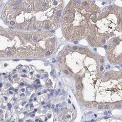
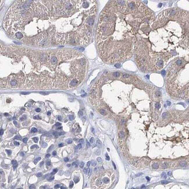
- Immunohistochemical staining of UGP2 in human kidney using UGP2 Polyclonal Antibody (Product # PA5-57019).
- Submitted by
- Invitrogen Antibodies (provider)
- Main image
- Experimental details
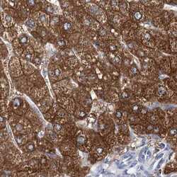
- Immunohistochemical staining of UGP2 in human liver using UGP2 Polyclonal Antibody (Product # PA5-57019).

- Submitted by
- Invitrogen Antibodies (provider)
- Main image
- Experimental details
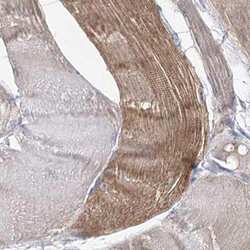
- Immunohistochemical staining of UGP2 in human skeletal muscle using UGP2 Polyclonal Antibody (Product # PA5-57019).

- Submitted by
- Invitrogen Antibodies (provider)
- Main image
- Experimental details
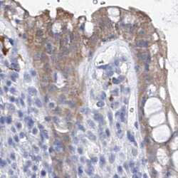
- Immunohistochemical staining of UGP2 in human rectum using UGP2 Polyclonal Antibody (Product # PA5-57019).

- Submitted by
- Invitrogen Antibodies (provider)
- Main image
- Experimental details
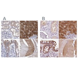
- Immunohistochemical staining of UGP2 in human kidney, liver, rectum and skeletal muscle using UGP2 Polyclonal Antibody (Product # PA5-57019) (A) shows similar protein distribution across tissues to an independent UGP2 Polyclonal Antibody (B).
