Explore
 Validate
 Learn
Learn Western blot
Antibody data
- Antibody Data
- Antigen structure
- References [0]
- Comments [0]
- Validations
- Western blot [3]
- Immunohistochemistry [1]
Submit
Validation data
Reference
Comment
Report error
- Product number
- PA5-27760 - Provider product page

- Provider
- Invitrogen Antibodies
- Product name
- UGP2 Polyclonal Antibody
- Antibody type
- Polyclonal
- Antigen
- Recombinant protein fragment
- Description
- Recommended positive controls: A431, H1299, HeLa, HepG2. Predicted reactivity: Mouse (98%), Rat (98%), Xenopus laevis (90%), Pig (98%), Chicken (93%), Rhesus Monkey (100%), Bovine (98%). Store product as a concentrated solution. Centrifuge briefly prior to opening the vial.
- Reactivity
- Human, Mouse, Rat
- Host
- Rabbit
- Isotype
- IgG
- Vial size
- 100 µL
- Concentration
- 0.56 mg/mL
- Storage
- Store at 4°C short term. For long term storage, store at -20°C, avoiding freeze/thaw cycles.
No comments: Submit comment
Supportive validation
- Submitted by
- Invitrogen Antibodies (provider)
- Main image
- Experimental details
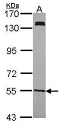
- Western Blot using UGP2 Polyclonal Antibody (Product # PA5-27760). Sample (30 µg of whole cell lysate). Lane A: H1299 . 7.5% SDS PAGE. UGP2 antibody . UGP2 Polyclonal Antibody (Product # PA5-27760) diluted at 1:1,000.

- Submitted by
- Invitrogen Antibodies (provider)
- Main image
- Experimental details
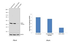
- Knockdown of UGP2 was achieved by transfecting Hep G2 cells with UGP2 specific siRNA (Silencer® select Product # S14645). Western blot analysis (Fig. a) was performed using whole cell extracts from the UGP2 knockdown cells (Lane 3), non-specific scrambled siRNA transfected cells (Lane 2) and untransfected cells (Lane 1). The blot was probed with UGP2 Polyclonal Antibody (Product # PA5-27760, 1:1000 dilution) and Goat anti-Rabbit IgG (H+L) Superclonal™ Secondary Antibody, HRP conjugate (Product # A27036, 1:4000 dilution). Densitometric analysis of this western blot is shown in histogram (Fig. b). Loss of signal upon siRNA mediated knock down confirms that antibody is specific to UGP2.

- Submitted by
- Invitrogen Antibodies (provider)
- Main image
- Experimental details
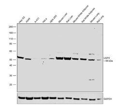
- Western blot was performed using Anti-UGP2 Polyclonal Antibody (Product # PA5-27760) and a 56 kDa band corresponding to UGP2 was observed across all the cell lines and tissues tested. Whole cell extracts (30 µg lysate) of Hep G2 (Lane 1), A549 (Lane 2), A-431 (Lane 3), HeLa (Lane 4), HEK-293 (Lane 5), tissue extracts of Mouse Liver (Lane 6), Rat Liver (Lane 7), Mouse White Adipose (Lane 8), Rat White Adipose (Lane 9), Mouse Lung (Lane 10) and Rat Lung (Lane 11) were electrophoresed using NuPAGE™ 4-12% Bis-Tris Protein Gel (Product # NP0322BOX). Resolved proteins were then transferred onto a nitrocellulose membrane (Product # IB23001) by iBlot® 2 Dry Blotting System (Product # IB21001). The blot was probed with the primary antibody (1:1000 dilution) and detected by chemiluminescence using Goat Anti-Rabbit IgG Secondary Antibody, HRP conjugate (Product # A27036, 1:4000 dilution) using the iBright FL 1000 (Product # A32752). Chemiluminescent detection was performed using Novex® ECL Chemiluminescent Substrate Reagent Kit (Product # WP20005).

Supportive validation
- Submitted by
- Invitrogen Antibodies (provider)
- Main image
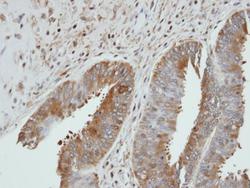
- Experimental details
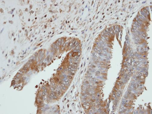
- Immunohistochemical analysis of paraffin-embedded UGP2 xenograft, using UGP2 (Product # PA5-27760) antibody at 1:100 dilution. Antigen Retrieval: EDTA based buffer, pH 8.0, 15 min.