 Explore
 Validate
 Learn
Learn Immunocytochemistry
Antibody data
- Antibody Data
- Antigen structure
- References [11]
- Comments [0]
- Validations
- Immunocytochemistry [1]
- Immunohistochemistry [1]
Submit
Validation data
Reference
Comment
Report error
- Product number
- HPA014791 - Provider product page

- Provider
- Atlas Antibodies
- Proper citation
- Atlas Antibodies Cat#HPA014791, RRID:AB_1847250
- Product name
- Anti-CLPTM1L
- Antibody type
- Polyclonal
- Description
- Polyclonal Antibody against Human CLPTM1L, Gene description: CLPTM1-like, Alternative Gene Names: CRR9, FLJ14400, Validated applications: IHC, ICC, Uniprot ID: Q96KA5, Storage: Store at +4°C for short term storage. Long time storage is recommended at -20°C.
- Reactivity
- Human
- Host
- Rabbit
- Conjugate
- Unconjugated
- Isotype
- IgG
- Vial size
- 100 µl
- Concentration
- 0.1 mg/ml
- Storage
- Store at +4°C for short term storage. Long time storage is recommended at -20°C.
- Handling
- The antibody solution should be gently mixed before use.
Submitted references CLPTM1L expression predicts recurrence of patients with intermediate‑ and high‑risk stage IB‑IIB cervical cancer undergoing radical hysterectomy followed by TP as adjuvant chemotherapy
CLPTM1L is a GPI-anchoring pathway component targeted by HCMV
Spatial proteomics reveals secretory pathway disturbances caused by neuropathy-associated TECPR2
Targeted biologic inhibition of both tumor cell-intrinsic and intercellular CLPTM1L/CRR9-mediated chemotherapeutic drug resistance
CLPTM1L Is a Novel Putative Oncogene Promoting Tumorigenesis in Oral Squamous Cell Carcinoma
CLPTM1L induces estrogen receptor β signaling-mediated radioresistance in non-small cell lung cancer cells
Mining the Plasma Cell Transcriptome for Novel Cell Surface Proteins
Development of primary human pancreatic cancer organoids, matched stromal and immune cells and 3D tumor microenvironment models
Novel Anti-CRR9/CLPTM1L Antibodies with Antitumorigenic Activity Inhibit Cell Surface Accumulation, PI3K Interaction, and Survival Signaling
Common variations in TERT-CLPTM1L locus are reproducibly associated with the risk of nasopharyngeal carcinoma in Chinese populations
CLPTM1L Promotes Growth and Enhances Aneuploidy in Pancreatic Cancer Cells
Awazu Y, Fukuda T, Noda T, Uchikura E, Nanno S, Imai K, Yamauchi M, Yasui T, Sumi T
Oncology Letters 2023;26(2)
Oncology Letters 2023;26(2)
CLPTM1L is a GPI-anchoring pathway component targeted by HCMV
Kol I, Rishiq A, Cohen M, Kahlon S, Pick O, Dassa L, Stein N, Bar-On Y, Wolf D, Seidel E, Mandelboim O
Journal of Cell Biology 2023;222(9)
Journal of Cell Biology 2023;222(9)
Spatial proteomics reveals secretory pathway disturbances caused by neuropathy-associated TECPR2
Nalbach K, Schifferer M, Bhattacharya D, Ho-Xuan H, Tseng W, Williams L, Stolz A, Lichtenthaler S, Elazar Z, Behrends C
Nature Communications 2023;14(1)
Nature Communications 2023;14(1)
Targeted biologic inhibition of both tumor cell-intrinsic and intercellular CLPTM1L/CRR9-mediated chemotherapeutic drug resistance
Parashar D, Geethadevi A, McAllister D, Ebben J, Peterson F, Jensen D, Bishop E, Pradeep S, Volkman B, Dwinell M, Chaluvally-Raghavan P, James M
npj Precision Oncology 2021;5(1)
npj Precision Oncology 2021;5(1)
CLPTM1L Is a Novel Putative Oncogene Promoting Tumorigenesis in Oral Squamous Cell Carcinoma
Hou Y, Xue F, Fu Y, Feng G, Wang R, Yuan H
Cell Transplantation 2021;30
Cell Transplantation 2021;30
CLPTM1L induces estrogen receptor β signaling-mediated radioresistance in non-small cell lung cancer cells
Li H, Che J, Jiang M, Cui M, Feng G, Dong J, Zhang S, Lu L, Liu W, Fan S
Cell Communication and Signaling 2020;18(1)
Cell Communication and Signaling 2020;18(1)
Mining the Plasma Cell Transcriptome for Novel Cell Surface Proteins
Trezise S, Karnowski A, Fedele P, Mithraprabhu S, Liao Y, D’Costa K, Kueh A, Hardy M, Owczarek C, Herold M, Spencer A, Shi W, Willis S, Nutt S, Corcoran L
International Journal of Molecular Sciences 2018;19(8):2161
International Journal of Molecular Sciences 2018;19(8):2161
Development of primary human pancreatic cancer organoids, matched stromal and immune cells and 3D tumor microenvironment models
Tsai S, McOlash L, Palen K, Johnson B, Duris C, Yang Q, Dwinell M, Hunt B, Evans D, Gershan J, James M
BMC Cancer 2018;18(1)
BMC Cancer 2018;18(1)
Novel Anti-CRR9/CLPTM1L Antibodies with Antitumorigenic Activity Inhibit Cell Surface Accumulation, PI3K Interaction, and Survival Signaling
Puskás L, Mán I, Szebeni G, Tiszlavicz L, Tsai S, James M
Molecular Cancer Therapeutics 2016;15(5):985-997
Molecular Cancer Therapeutics 2016;15(5):985-997
Common variations in TERT-CLPTM1L locus are reproducibly associated with the risk of nasopharyngeal carcinoma in Chinese populations
Zhang Y, Zhang X, Zhang H, Zhai Y, Wang Z, Li P, Yu L, Xia X, Zhang Y, Zeng Y, He F, Zhou G
Oncotarget 2015;7(1):759-770
Oncotarget 2015;7(1):759-770
CLPTM1L Promotes Growth and Enhances Aneuploidy in Pancreatic Cancer Cells
Jia J, Bosley A, Thompson A, Hoskins J, Cheuk A, Collins I, Parikh H, Xiao Z, Ylaya K, Dzyadyk M, Cozen W, Hernandez B, Lynch C, Loncarek J, Altekruse S, Zhang L, Westlake C, Factor V, Thorgeirsson S, Bamlet W, Hewitt S, Petersen G, Andresson T, Amundadottir L
Cancer Research 2014;74(10):2785-2795
Cancer Research 2014;74(10):2785-2795
No comments: Submit comment
Supportive validation
- Submitted by
- Atlas Antibodies (provider)
- Main image
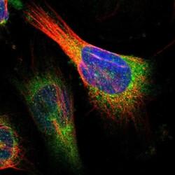
- Experimental details
- Immunofluorescent staining of human cell line U-251 MG shows localization to endoplasmic reticulum.
- Sample type
- Human

Supportive validation
- Submitted by
- Atlas Antibodies (provider)
- Enhanced method
- Orthogonal validation
- Main image
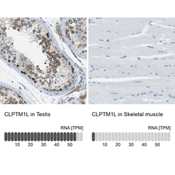
- Experimental details
- Immunohistochemistry analysis in human testis and skeletal muscle tissues using HPA014791 antibody. Corresponding CLPTM1L RNA-seq data are presented for the same tissues.
- Sample type
- Human
- Protocol
- Protocol
