 Explore
 Validate
 Learn
Learn Western blot
 ELISA
ELISAAntibody data
- Antibody Data
- Antigen structure
- References [2]
- Comments [0]
- Validations
- Western blot [1]
- Immunocytochemistry [1]
- Immunohistochemistry [1]
- Flow cytometry [1]
Submit
Validation data
Reference
Comment
Report error
- Product number
- ABIN952441 - Provider product page

- Provider
- antibodies-online
- Product name
- anti-Galactosamine (N-Acetyl)-6-Sulfate Sulfatase (GALNS) (Center) antibody
- Antibody type
- Polyclonal
- Antigen
- Other
- Reactivity
- Human
- Host
- Rabbit
- Vial size
- 0.1 mg
Submitted references Descriptively quantitative relationship between mutated N-acetylgalactosamine-6-sulfatase and mucopolysaccharidosis IVA.
Role of macrophage oxidative burst in the action of anthrax lethal toxin.
Yan S, Wu G
Biopolymers 2009;92(5):399-404
Biopolymers 2009;92(5):399-404
Role of macrophage oxidative burst in the action of anthrax lethal toxin.
Hanna PC, Kruskal BA, Ezekowitz RA, Bloom BR, Collier RJ
Molecular medicine (Cambridge, Mass.) 1994 Nov;1(1):7-18
Molecular medicine (Cambridge, Mass.) 1994 Nov;1(1):7-18
No comments: Submit comment
Supportive validation
- Submitted by
- antibodies-online (provider)
- Main image
- Experimental details
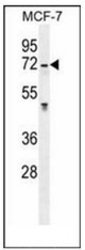
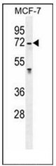
- Western blot analysis of GALNS Antibody (Center) Cat.-No AP51765PU-N in MCF-7 cell line lysates (35ug/lane). This demonstrates the GALNS antibody detected the GALNS protein (arrow).
Supportive validation
- Submitted by
- antibodies-online (provider)
- Main image

- Experimental details
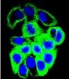
- Confocal immunofluorescent analysis of GALNS Antibody (Center) Cat.-No AP51765PU-N with MCF-7 cell followed by Alexa Fluor 488-conjugated goat anti-rabbit lgG (green).DAPI was used to stain the cell nuclear (blue).
Supportive validation
- Submitted by
- antibodies-online (provider)
- Main image

- Experimental details
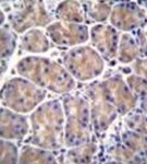
- Immunohistochemistry analysis in formalin fixed and paraffin embedded human stomach tissue reacted with GALNS Antibody (Center) Cat.-No AP51765PU-N followed by peroxidase conjugation of the secondary antibody and DAB staining.
Supportive validation
- Submitted by
- antibodies-online (provider)
- Main image
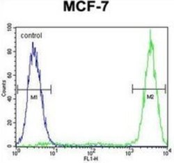
- Experimental details
- Flow cytometric analysis of MCF-7 cells using GALNS Antibody (Center) Cat.-No AP51765PU-N (right histogram) compared to a negative control cell (left histogram).FITC-conjugated goat-anti-rabbit secondary antibodies were used for the analysis.
