Explore
 Validate
 Learn
Learn Western blot
 Immunocytochemistry
Antibody data
- Antibody Data
- Antigen structure
- References [0]
- Comments [0]
- Validations
- Immunocytochemistry [2]
- Immunoprecipitation [1]
Submit
Validation data
Reference
Comment
Report error
- Product number
- PA5-112217 - Provider product page

- Provider
- Invitrogen Antibodies
- Product name
- UTP18 Polyclonal Antibody
- Antibody type
- Polyclonal
- Antigen
- Other
- Reactivity
- Human
- Host
- Rabbit
- Isotype
- IgG
- Vial size
- 100 μL
- Concentration
- 1 mg/mL
- Storage
- Store at 4°C short term. For long term storage, store at -20°C, avoiding freeze/thaw cycles.
No comments: Submit comment
Supportive validation
- Submitted by
- Invitrogen Antibodies (provider)
- Main image
- Experimental details
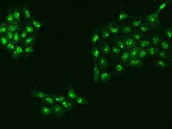
- Immunocytochemistry-Immunofluorescence analysis of UTP18 in A431 cells. Cells were fixed with 4% PFA, permeabilized with 0.1% Triton X-100 in PBS, blocked with 10% serum, and incubated with UTP18 Polyclonal Antibody (Product # PA5-112217) (1:200) at 4°C overnight. Then cells were stained with the Alexa Fluor 488-conjugated Goat Anti-rabbit IgG secondary antibody (green). Positive staining was localized to Nucleus.
- Submitted by
- Invitrogen Antibodies (provider)
- Main image
- Experimental details
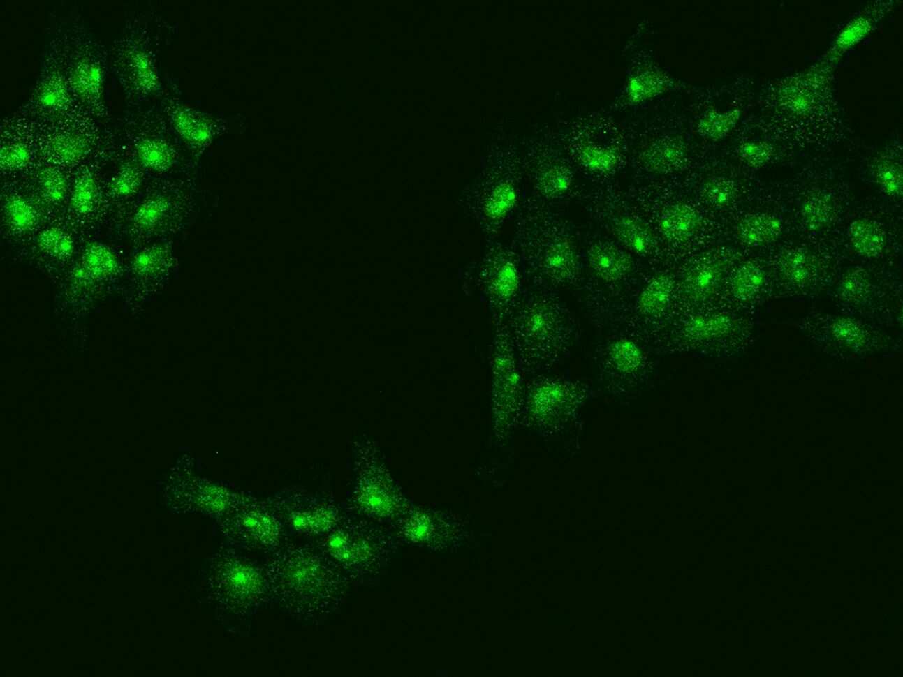
- Immunocytochemistry-Immunofluorescence analysis of UTP18 in A431 cells. Cells were fixed with 4% PFA, permeabilized with 0.1% Triton X-100 in PBS, blocked with 10% serum, and incubated with UTP18 Polyclonal Antibody (Product # PA5-112217) (1:200) at 4°C overnight. Then cells were stained with the Alexa Fluor 488-conjugated Goat Anti-rabbit IgG secondary antibody (green). Positive staining was localized to Nucleus.

Supportive validation
- Submitted by
- Invitrogen Antibodies (provider)
- Main image
- Experimental details
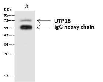
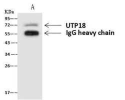
- Immunoprecipitation of UTP18 was performed on (Lane A) 0.5 mg U-251MG whole cell lysate using 4 µL of UTP18 Polyclonal Antibody (Product # PA5-112217) at a dilution of 1:100, and 60 µg of Immunomagnetic beads Protein A/G. A Goat Anti-Rabbit IgG (H+L)/HRP was used as a secondary antibody at a dilution of 1:10,000. Developed using the ECL technique and performed under reducing conditions. Predicted band size: 62 kDa. Observed band size: 70 kDa.