Explore
 Validate
 Learn
Learn Western blot
 Immunocytochemistry
Antibody data
- Antibody Data
- Antigen structure
- References [1]
- Comments [0]
- Validations
- Immunocytochemistry [1]
Submit
Validation data
Reference
Comment
Report error
- Product number
- HPA052378 - Provider product page

- Provider
- Atlas Antibodies
- Proper citation
- Atlas Antibodies Cat#HPA052378, RRID:AB_2681805
- Product name
- Anti-UTP18
- Antibody type
- Polyclonal
- Description
- Polyclonal Antibody against Human UTP18, Gene description: UTP18 small subunit (SSU) processome component homolog (yeast), Alternative Gene Names: CGI-48, WDR50, Validated applications: WB, IHC, ICC, Uniprot ID: Q9Y5J1, Storage: Store at +4°C for short term storage. Long time storage is recommended at -20°C.
- Reactivity
- Human
- Host
- Rabbit
- Conjugate
- Unconjugated
- Isotype
- IgG
- Vial size
- 100 µl
- Concentration
- 0.05 mg/ml
- Storage
- Store at +4°C for short term storage. Long time storage is recommended at -20°C.
- Handling
- The antibody solution should be gently mixed before use.
Submitted references A small subunit processome protein promotes cancer by altering translation
Yang H, Kim T, Song S, Menon L, Jiang X, Huang W, Black P, Park P, Carroll R, Johnson M
Oncogene 2014;34(34):4471-4481
Oncogene 2014;34(34):4471-4481
No comments: Submit comment
Supportive validation
- Submitted by
- Atlas Antibodies (provider)
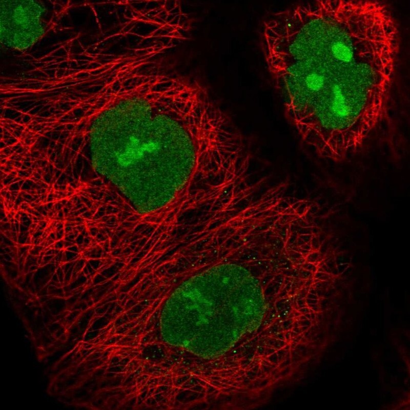
- Main image
- Experimental details
- Immunofluorescent staining of human cell line A-431 shows localization to nucleus & nucleoli.
- Sample type
- Human