Explore
 Validate
 Learn
Learn Western blot
 Immunocytochemistry
Antibody data
- Antibody Data
- Antigen structure
- References [2]
- Comments [0]
- Validations
- Immunocytochemistry [2]
- Other assay [2]
Submit
Validation data
Reference
Comment
Report error
- Product number
- PA5-45768 - Provider product page

- Provider
- Invitrogen Antibodies
- Product name
- FOLR2 Polyclonal Antibody
- Antibody type
- Polyclonal
- Antigen
- Synthetic peptide
- Description
- Peptide sequence: LCEGLWSHSY KVSNYSRGSG RCIQMWFDSA QGNPNEEVAR FYAAAMHVNA Sequence homology: Cow: 100%; Dog: 100%; Guinea Pig: 86%; Horse: 100%; Human: 100%; Mouse: 100%; Pig: 93%; Rabbit: 93%; Rat: 100%
- Reactivity
- Human
- Host
- Rabbit
- Isotype
- IgG
- Vial size
- 100 μL
- Concentration
- 0.5 mg/mL
- Storage
- -20°C, Avoid Freeze/Thaw Cycles
Submitted references Cancer Selective Target Degradation by Folate-Caged PROTACs.
Bortezomib-Loaded Mesoporous Silica Nanoparticles Selectively Alter Metabolism and Induce Death in Multiple Myeloma Cells.
Liu J, Chen H, Liu Y, Shen Y, Meng F, Kaniskan HÜ, Jin J, Wei W
Journal of the American Chemical Society 2021 May 19;143(19):7380-7387
Journal of the American Chemical Society 2021 May 19;143(19):7380-7387
Bortezomib-Loaded Mesoporous Silica Nanoparticles Selectively Alter Metabolism and Induce Death in Multiple Myeloma Cells.
Nigro A, Frattaruolo L, Fava M, De Napoli I, Greco M, Comandè A, De Santo M, Pellegrino M, Ricci E, Giordano F, Perrotta I, Leggio A, Pasqua L, Sisci D, Cappello AR, Morelli C
Cancers 2020 Sep 21;12(9)
Cancers 2020 Sep 21;12(9)
No comments: Submit comment
Supportive validation
- Submitted by
- Invitrogen Antibodies (provider)
- Main image
- Experimental details
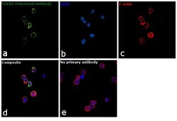
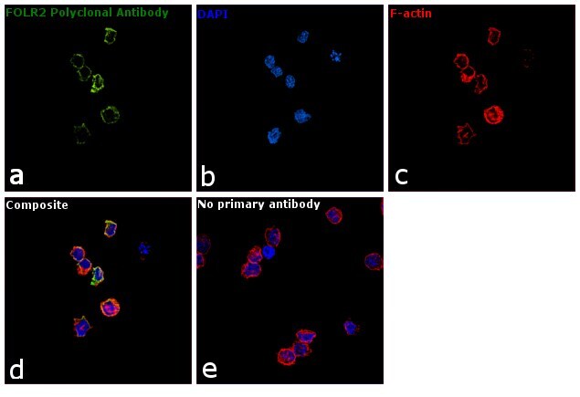
- Immunofluorescence analysis of FOLR2 was performed using 70% confluent log phase Raji cells. The cells were fixed with 4% paraformaldehyde for 10 minutes, permeabilized with 0.1% Triton™ X-100 for 15 minutes, and blocked with 1% BSA for 1 hour at room temperature. The cells were labeled with FOLR2 Polyclonal Antibody (Product # PA5-45768) at 5 µg/mL in 0.1% BSA, incubated at 4 degree Celsius overnight and then labeled with Goat anti-Rabbit IgG (H+L) Superclonal™ Secondary Antibody, Alexa Fluor® 488 conjugate (Product # A27034) at a dilution of 1:2000 for 45 minutes at room temperature (Panel a: green). Nuclei (Panel b: blue) were stained with SlowFade® Gold Antifade Mountant with DAPI (Product # S36938). F-actin (Panel c: red) was stained with Rhodamine Phalloidin (Product # R415, 1:300). Panel d represents the merged image showing cell membrane localization. Panel e represents control cells with no primary antibody to assess background. The images were captured at 60X magnification.
- Submitted by
- Invitrogen Antibodies (provider)
- Main image

- Experimental details
- Immunofluorescence analysis of FOLR2 was performed using 70% confluent log phase Raji cells. The cells were fixed with 4% paraformaldehyde for 10 minutes, permeabilized with 0.1% Triton™ X-100 for 15 minutes, and blocked with 1% BSA for 1 hour at room temperature. The cells were labeled with FOLR2 Polyclonal Antibody (Product # PA5-45768) at 5 µg/mL in 0.1% BSA, incubated at 4 degree Celsius overnight and then labeled with Goat anti-Rabbit IgG (Heavy Chain) Superclonal™ Secondary Antibody, Alexa Fluor® 488 conjugate (Product # A27034) at a dilution of 1:2000 for 45 minutes at room temperature (Panel a: green). Nuclei (Panel b: blue) were stained with SlowFade® Gold Antifade Mountant with DAPI (Product # S36938). F-actin (Panel c: red) was stained with Rhodamine Phalloidin (Product # R415, 1:300). Panel d represents the merged image showing cell membrane localization. Panel e represents control cells with no primary antibody to assess background. The images were captured at 60X magnification.

Supportive validation
- Submitted by
- Invitrogen Antibodies (provider)
- Main image
- Experimental details
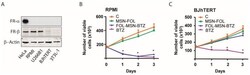
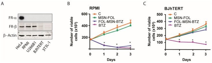
- Figure 1 FOL-MSN-BTZ kills FR+ MM cells but not normal cells. ( A ) 50 mug of non-denatured proteins (for FR-alpha detection) and denatured proteins (for FR-beta detection) from total lysates of indicated cell lines were loaded and subjected to WB analysis. beta-actin was used as loading control. FOL-MSN-BTZ induces death in FR+ RPMI-8226 (RPMI) MM cells ( B ) but not in FR- BJhTERT normal cells ( C ). Cells were treated for 1h with FOL-MSN-BTZ or left untreated (C = control). MSN-FOL was used as negative control and the free drug BTZ as positive control. (*) p < 0.05 vs. control.
- Submitted by
- Invitrogen Antibodies (provider)
- Main image
- Experimental details
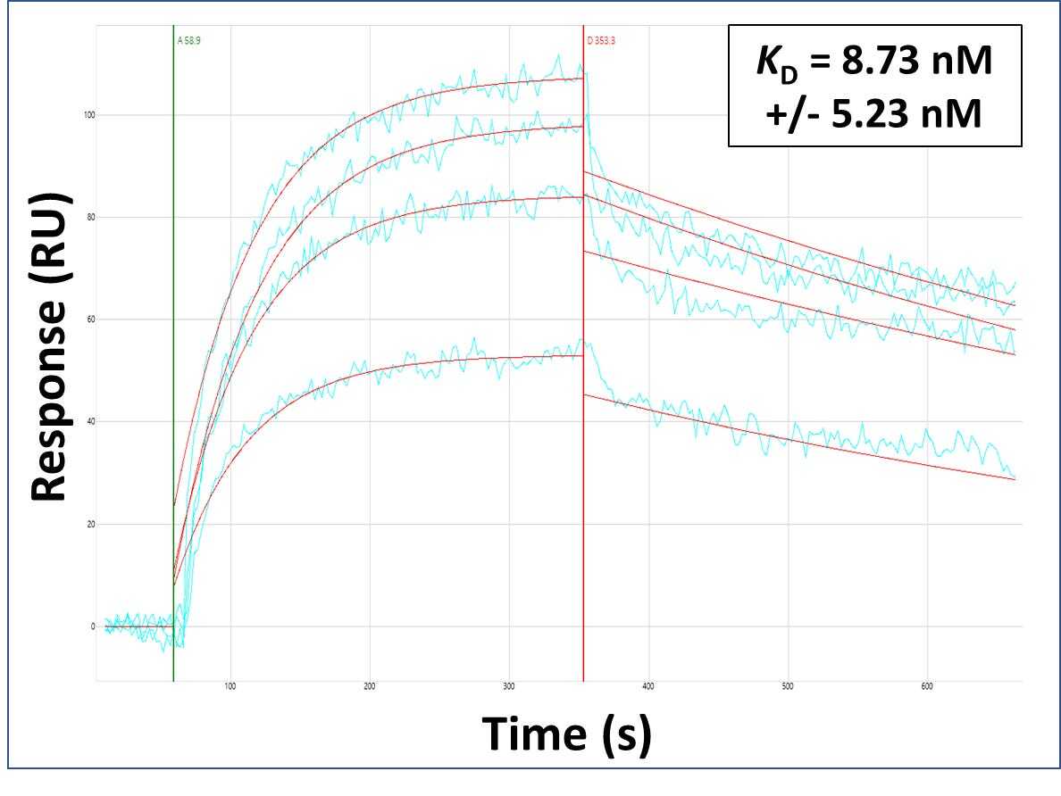
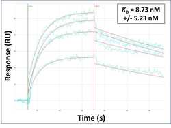
- Surface Plasmon Resonance of FOLR2 polyclonal antibody (Product # PA5-45768). Purified polyclonal antibodies were immobilized on a Protein A/G coated Carterra LSA sensor chip at concentrations of 5, and 50 µg/mL in duplicate. Antibodies on the surface were exposed to interaction with peptides sequentially via microfluidic controlled flow at 333 nm peptide concentration for kinetic characterization of the binders for affinity and specificity, followed by curve fitting using the Kinetics software. Kd determinations for both concentrations were averaged and results and standard deviation are shown.