Explore
 Validate
 Learn
Learn Western blot
Antibody data
- Antibody Data
- Antigen structure
- References [0]
- Comments [0]
- Validations
- Western blot [1]
- Immunocytochemistry [1]
Submit
Validation data
Reference
Comment
Report error
- Product number
- MAB5440 - Provider product page

- Provider
- R&D Systems
- Product name
- Mouse Semaphorin 3B Antibody
- Antibody type
- Monoclonal
- Description
- Protein A or G purified from hybridoma culture supernatant. Detects mouse Semaphorin 3B in direct ELISAs.
- Reactivity
- Mouse
- Host
- Rat
- Conjugate
- Unconjugated
- Antigen sequence
Q62177- Isotype
- IgG
- Antibody clone number
- 904201
- Vial size
- 100 ug
- Storage
- Use a manual defrost freezer and avoid repeated freeze-thaw cycles. 12 months from date of receipt, -20 to -70 °C as supplied. 1 month, 2 to 8 °C under sterile conditions after reconstitution. 6 months, -20 to -70 °C under sterile conditions after reconstitution.
No comments: Submit comment
Supportive validation
- Submitted by
- R&D Systems (provider)
- Main image
- Experimental details
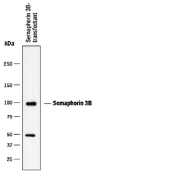
- Detection of Mouse Semaphorin 3B by Western Blot. Western blot shows lysates of NS0 mouse myeloma cell line transfected with mouse Semaphorin 3B. PVDF membrane was probed with 0.5 µg/mL of Rat Anti-Mouse Semaphorin 3B Monoclonal Antibody (Catalog # MAB5440) followed by HRP-conjugated Anti-Rat IgG Secondary Antibody (Catalog # HAF005). A specific band was detected for Semaphorin 3B at approximately 100 kDa (as indicated). This experiment was conducted under reducing conditions and using Immunoblot Buffer Group 1.
Supportive validation
- Submitted by
- R&D Systems (provider)
- Main image
- Experimental details
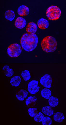
- Semaphorin 3B in NS0 Mouse Cell Line. Semaphorin 3B was detected in immersion fixed NS0 mouse myeloma cell line transfected with mouse Semaphorin 3B (upper panel) or irrelevant transfectant (lower panel) using Rat Anti-Mouse Semaphorin 3B Monoclonal Antibody (Catalog # MAB5440) at 10 µg/mL for 3 hours at room temperature. Cells were stained using the NorthernLights™ 557-conjugated Anti-Rat IgG Secondary Antibody (red; Catalog # NL013) and counterstained with DAPI (blue). Specific staining was localized to cytoplasm. View our protocol for Fluorescent ICC Staining of Non-adherent Cells.