Explore
 Validate
 Learn
Learn Western blot
Antibody data
- Antibody Data
- Antigen structure
- References [0]
- Comments [0]
- Validations
- Western blot [1]
- Immunocytochemistry [1]
- Immunohistochemistry [1]
Submit
Validation data
Reference
Comment
Report error
- Product number
- AF5389 - Provider product page

- Provider
- R&D Systems
- Product name
- Human/Mouse/Rat SHIP2 Antibody
- Antibody type
- Polyclonal
- Description
- Antigen Affinity-purified. Detects human, mouse and rat SHIP2 in Western blots.
- Reactivity
- Human, Mouse, Rat
- Host
- Sheep
- Conjugate
- Unconjugated
- Antigen sequence
O15357- Isotype
- IgG
- Vial size
- 100 ug
- Concentration
- LYOPH
- Storage
- Use a manual defrost freezer and avoid repeated freeze-thaw cycles. 12 months from date of receipt, -20 to -70 °C as supplied. 1 month, 2 to 8 °C under sterile conditions after reconstitution. 6 months, -20 to -70 °C under sterile conditions after reconstitution.
No comments: Submit comment
Supportive validation
- Submitted by
- R&D Systems (provider)
- Main image
- Experimental details
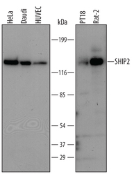
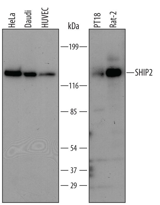
- Detection of Human/Mouse/Rat SHIP2 by Western Blot. Western blot shows lysates of HeLa human cervical epithelial carcinoma cell line, Daudi human Burkitt's lymphoma cell line, HUVEC human umbilical vein endothelial cells, PT18 mouse mast/basophil cell line, and Rat-2 rat embryonic fibroblast cell line. PVDF membrane was probed with 1 µg/mL of Sheep Anti-Human/Mouse/Rat SHIP2 Antigen Affinity-purified Polyclonal Antibody (Catalog # AF5389) followed by HRP-conjugated Anti-Sheep IgG Secondary Antibody (Catalog # HAF016). A specific band was detected for SHIP2 at approximately 140 kDa (as indicated). This experiment was conducted using Immunoblot Buffer Group 1.
Supportive validation
- Submitted by
- R&D Systems (provider)
- Main image

- Experimental details
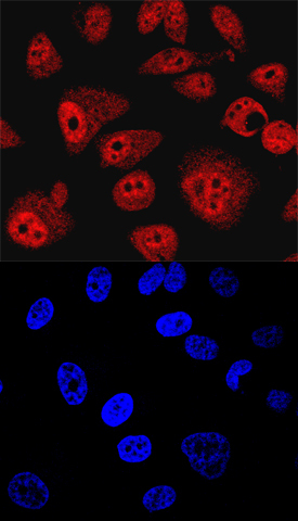
- SHIP2 in PC-3 Human Cell Line. SHIP2 was detected in immersion fixed PC-3 human prostate cancer cell line using Sheep Anti-Human/Mouse/Rat SHIP2 Antigen Affinity-purified Polyclonal Antibody (Catalog # AF5389) at 10 µg/mL for 3 hours at room temperature. Cells were stained using the NorthernLights™ 557-conjugated Anti-Sheep IgG Secondary Antibody (red, upper panel; Catalog # NL010) and counterstained with DAPI (blue, lower panel). Specific staining was localized to nuclei and cytoplasm. View our protocol for Fluorescent ICC Staining of Cells on Coverslips. This application has not yet been tested in mouse or rat samples.
Supportive validation
- Submitted by
- R&D Systems (provider)
- Main image
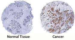
- Experimental details
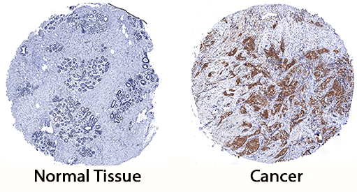
- SHIP2 in Human Breast Cancer Tissue. SHIP2 was detected in immersion fixed paraffin-embedded sections of human breast cancer tissue using Sheep Anti-Human/Mouse/Rat SHIP2 Antigen Affinity-purified Polyclonal Antibody (Catalog # AF5389) at 3 µg/mL for 1 hour at room temperature followed by incubation with the Anti-Goat IgG VisUCyte™ HRP Polymer Antibody (Catalog # VC004). Tissue was stained using DAB (brown) and counterstained with hematoxylin (blue). Specific staining was localized to cytoplasm in cancer cells. View our protocol for IHC Staining with VisUCyte HRP Polymer Detection Reagents.