Explore
 Validate
 Learn
Learn Immunocytochemistry
Antibody data
- Antibody Data
- Antigen structure
- References [1]
- Comments [0]
- Validations
- Immunocytochemistry [2]
- Immunohistochemistry [6]
- Other assay [2]
Submit
Validation data
Reference
Comment
Report error
- Product number
- PA5-59390 - Provider product page

- Provider
- Invitrogen Antibodies
- Product name
- UBE3B Polyclonal Antibody
- Antibody type
- Polyclonal
- Antigen
- Recombinant protein fragment
- Description
- Immunogen sequence: ANIMGHLNQH GFYSVLQILL TRGLARPRPC LSKGTLTAAF SLALRPVIAA QFSDNLIRPF LIHIMSVPAL VTHLSTVTPE RLTVLESHDM LRKFIIFLRD QDRCRDV Highest antigen sequence identity to the following orthologs: Mouse - 87%, Rat - 85%.
- Reactivity
- Human
- Host
- Rabbit
- Isotype
- IgG
- Vial size
- 100 μL
- Concentration
- 0.3 mg/mL
- Storage
- Store at 4°C short term. For long term storage, store at -20°C, avoiding freeze/thaw cycles.
Submitted references TRIB3 promotes MYC-associated lymphoma development through suppression of UBE3B-mediated MYC degradation.
Li K, Wang F, Yang ZN, Zhang TT, Yuan YF, Zhao CX, Yeerjiang Z, Cui B, Hua F, Lv XX, Zhang XW, Yu JJ, Liu SS, Yu JM, Shang S, Xiao Y, Hu ZW
Nature communications 2020 Dec 9;11(1):6316
Nature communications 2020 Dec 9;11(1):6316
No comments: Submit comment
Supportive validation
- Submitted by
- Invitrogen Antibodies (provider)
- Main image
- Experimental details
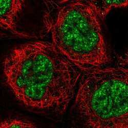
- Immunofluorescent staining of UBE3B in human cell line A-431 shows positivity in nucleus but excluded from the nucleoli. Samples were probed using an UBE3B Polyclonal Antibody (Product # PA5-59390).

- Submitted by
- Invitrogen Antibodies (provider)
- Main image

- Experimental details
- Immunofluorecent analysis of UBE3B in human cell line A-431 using UBE3B Polyclonal Antibody (Product # PA5-59390). Staining shows localization to nuclear speckles.

Supportive validation
- Submitted by
- Invitrogen Antibodies (provider)
- Main image
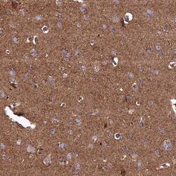
- Experimental details
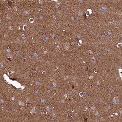
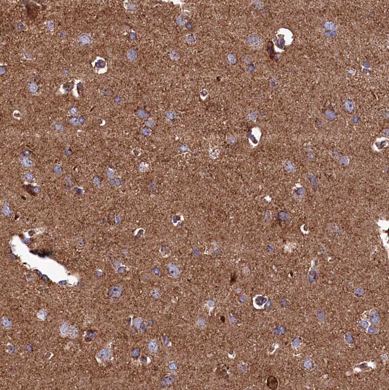
- Immunohistochemical analysis of UBE3B in human cerebral cortex using UBE3B Polyclonal Antibody (Product # PA5-59390) shows strong positivity in neuropil.
- Submitted by
- Invitrogen Antibodies (provider)
- Main image
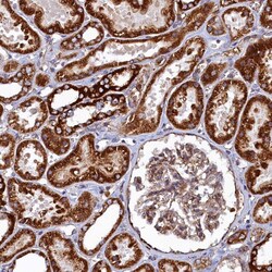
- Experimental details
- Immunohistochemical analysis of UBE3B in human kidney using UBE3B Polyclonal Antibody (Product # PA5-59390) shows strong positivity in cytoplasm in cells in tubules.

- Submitted by
- Invitrogen Antibodies (provider)
- Main image
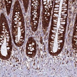
- Experimental details
- Immunohistochemical analysis of UBE3B in human rectum using UBE3B Polyclonal Antibody (Product # PA5-59390) shows strong positivity in cytoplasm in glandular cells.

- Submitted by
- Invitrogen Antibodies (provider)
- Main image
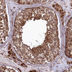
- Experimental details
- Immunohistochemical analysis of UBE3B in human testis using UBE3B Polyclonal Antibody (Product # PA5-59390) shows strong positivity in cytoplasm in cells in seminiferous ducts.

- Submitted by
- Invitrogen Antibodies (provider)
- Main image
- Experimental details
- Immunohistochemical analysis of UBE3B in human cerebral cortex using UBE3B Polyclonal Antibody (Product # PA5-59390) shows strong positivity in neuropil.

- Submitted by
- Invitrogen Antibodies (provider)
- Main image

- Experimental details
- Immunohistochemical analysis of UBE3B in human testis using UBE3B Polyclonal Antibody (Product # PA5-59390) shows strong positivity in cytoplasm in cells in seminiferous ducts.

Supportive validation
- Submitted by
- Invitrogen Antibodies (provider)
- Main image
- Experimental details
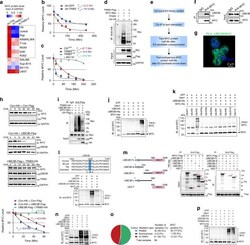
- Fig. 3 TRIB3 supports MYC stability by disturbing UBE3B-mediated MYC ubiquitination. a Heatmap presenting TRIB3 expression in 11 blood cancer cell lines and 1 primary human lymphoma cell line (T144) with or without TRIB3 depletion. b Effect of TRIB3 overexpression on MYC degradation in vivo. Raji cells were infected with TRIB3 - or GFP - adenovirus, and 12 h later, incubated with cycloheximide (CHX; 10 mug/mL) for the indicated times. The data are presented as the means +- S.E.M from three independent experiments. c Effect of TRIB3 deletion on MYC degradation in vivo. Control or TRIB3 -deleted Raji cells were incubated with CHX (10 mug/mL) or CHX plus MG132 (5 mum) for the indicated times. The MYC protein was detected by western blot; GAPDH was used as a loading control. The data are presented as the means +- S.E.M from three independent experiments. d Effect of TRIB3 overexpression on MYC ubiquitination in vivo. HEK 293T cells were transfected with the indicated plasmids for 24 h. Extracts of cells were IP with an anti-HA Ab. Ubiquitinated MYC was detected by immunoblotting. The data are presented as presentative from three independent experiments. e Strategy for screening the potential E3 ligases of MYC. f The interaction of UBE3B and MYC was evaluated by Co-IP assays. Raji cell extracts were IP with rabbit immunoglobulin G (IgG) or an anti-MYC Ab and blotted with an anti-UBE3B Ab. The data are presented as presentative from three independent experiments. g Colocalization o

- Submitted by
- Invitrogen Antibodies (provider)
- Main image
- Experimental details
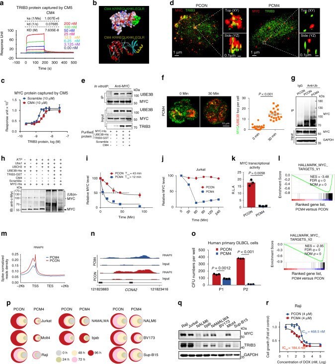
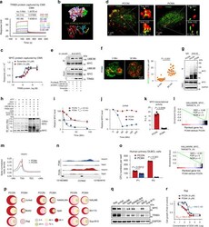
- Fig. 6 Disturbing the TRIB3/MYC interaction destabilizes MYC and inhibits lymphoma. a The kinetic interaction of CM4 and TRIB3 was determined by surface plasmon resonance (SPR) analyses. b The highest scoring Dock model of the CM4 and TRIB3 complex is shown. Upper: the surface of CM4 (green) and the TRIB3 complex. Below: the 3D structure of CM4 (yellow) and the TRIB3 complex. c The kinetic interaction of the TRIB3 and MYC proteins was determined by SPR analyses with or without CM4. d Structured illumination microscopic (SIM) images of PCON- or PCM4-treated Raji cells (30 min) stained for MYC and TRIB3. Dotted line border square: area processed for 3D surface rendering (insets). e In vitro interaction assays show that the CM4 peptide increased the binding of MYC and UBE3B. f Representative images of MYC and UBE3B foci (left) and quantification of the number of MYC/UBE3B colocalized foci in Raji cells before and after PCM4 treatment. Scale bar, 10 mum. Data are represented as means +- SEM. Statistical significance was determined by two-tailed Student's t test. P value: 3.74 x 10 -13 . g Effect of PCM4 on MYC ubiquitination. Extracts of PCON- and PCM4-treated Raji cells were IP with an anti-Ub Ab. Ubiquitinated MYC was detected by immunoblotting. h In vitro ubiquitination assays show that the CM4 peptide restored the polyubiquitination of MYC. i Effect of PCM4 on MYC protein degradation. Raji cells were treated with CHX (10 mug/mL) and the PCON or PCM4 peptide (4 mum) for the in