Explore
 Validate
 Learn
Learn Western blot
 Immunohistochemistry
Antibody data
- Antibody Data
- Antigen structure
- References [1]
- Comments [0]
- Validations
- Immunohistochemistry [4]
- Other assay [1]
Submit
Validation data
Reference
Comment
Report error
- Product number
- MA5-16229 - Provider product page

- Provider
- Invitrogen Antibodies
- Product name
- IL-17F Monoclonal Antibody (4H1629.1)
- Antibody type
- Monoclonal
- Antigen
- Recombinant full-length protein
- Description
- The human IL17F protein shows 60% sequence similarity with Rat and 57% with Mouse. Does not react with Rat or Mouse recombinant IL17F proteins. Immunogen sequence: MRKIPKVGHT FFQKPESCPP VPGGSMKLDI GIINENQRVS MSRNIESRST SPWNYTVTWD PNRYPSEVVQ AQCRNLGCIN AQGKEDISMN SVPIQQETLV VRRKHQGCSV SFQLEKVLVT VGCTCVTPVI HHVQ
- Reactivity
- Human
- Host
- Mouse
- Isotype
- IgG
- Antibody clone number
- 4H1629.1
- Vial size
- 100 μg
- Concentration
- 1.0 mg/mL
- Storage
- Store at 4°C short term. For long term storage, store at -20°C, avoiding freeze/thaw cycles.
Submitted references Exaggerated IL-17A activity in human in vivo recall responses discriminates active tuberculosis from latent infection and cured disease.
Pollara G, Turner CT, Rosenheim J, Chandran A, Bell LCK, Khan A, Patel A, Peralta LF, Folino A, Akarca A, Venturini C, Baker T, Ecker S, Ricciardolo FLM, Marafioti T, Ugarte-Gil C, Moore DAJ, Chain BM, Tomlinson GS, Noursadeghi M
Science translational medicine 2021 May 5;13(592)
Science translational medicine 2021 May 5;13(592)
No comments: Submit comment
Supportive validation
- Submitted by
- Invitrogen Antibodies (provider)
- Main image
- Experimental details
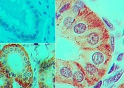
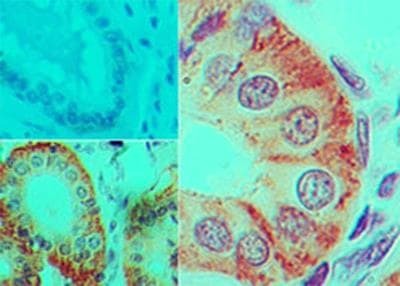
- Immunohistochemical analysis of IL-17F in formalin-fixed paraffin-embedded human colon tissue. Samples were incubated in IL-17F monoclonal antibody (Product # MA5-16229) using a dilution of 5 µg/mL. Isotype control (top left) and IL-17F antibody (bottom left and right).
- Submitted by
- Invitrogen Antibodies (provider)
- Main image
- Experimental details
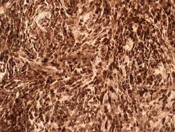
- Immunohistochemical analysis of IL-17F in a tissue section of human lymph node cancer. Samples were incubated in IL-17F monoclonal antibody (Product # MA5-16229) using a dilution of 5 µg/mL.
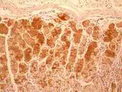
- Submitted by
- Invitrogen Antibodies (provider)
- Main image
- Experimental details
- Immunohistochemical analysis of IL-17F in a tissue section of human stomach cancer. Samples were incubated in IL-17F monoclonal antibody (Product # MA5-16229) using a dilution of 5 µg/mL. Diffused cytoplasmic staining was observed in the tissue section and interestingly, the expression levels in cancerous tissue were found to be weaker than the parallel stained normal tissue section.

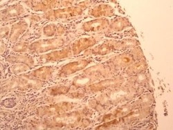
- Submitted by
- Invitrogen Antibodies (provider)
- Main image
- Experimental details
- Immunohistochemical analysis of IL-17F in a tissue section of normal human stomach. Samples were incubated in IL-17F monoclonal antibody (Product # MA5-16229) using a dilution of 5 µg/mL. Cytoplasmic expression was observed all over the tissue section and the staining was found to be more intense expecially in the chief cells of the glands.

Supportive validation
- Submitted by
- Invitrogen Antibodies (provider)
- Main image

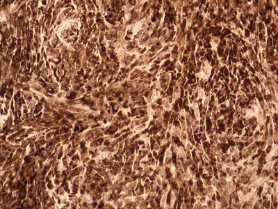
- Experimental details
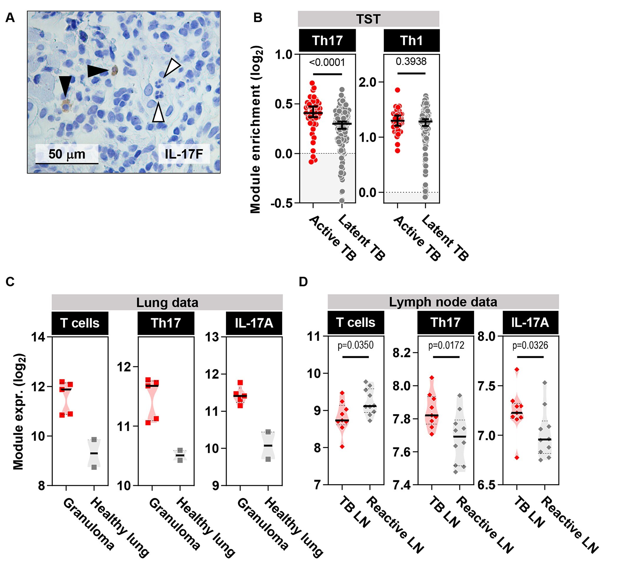
- Figure 4 Active TB is characterized by elevated Th17 cells at the site of TST. (A) IL-17F immunohistochemistry (brown) in TST of patients with active TB. Black arrowheads point to mononuclear cells that express IL-17F and white arrowheads point to polymorphonuclear cells that do not express IL-17F. (B) Th17 and Th1 cell module enrichment in TST samples relative to saline injection in patients with active or latent TB. Analyses were performed on samples from 48 and 191 participants with active and latent TB, respectively. Horizontal lines and error bars on scatter dot plots represent medians with 95% confidence interval. (C and D) Expression of T cells, Th17 cells, and IL-17A bioactivity modules from the site of human TB granulomata (n=5) relative to healthy lung tissue (n=2) (dataset GSE20050) (C) and in human Mtb-infected lymph nodes (LN) (n=9) relative to reactive lymph nodes that do not display evidence of granulomatous inflammation or cancer (n=10) (D) (dataset E-MTAB-2547). Violin plots represent frequency distribution of all samples, with bold lines representing median values. All p values were calculated by Mann-Whitney tests.