Explore
 Validate
 Learn
Learn Western blot
 Immunocytochemistry
Antibody data
- Antibody Data
- Antigen structure
- References [1]
- Comments [0]
- Validations
- Immunocytochemistry [1]
- Other assay [1]
Submit
Validation data
Reference
Comment
Report error
- Product number
- MA5-24702 - Provider product page

- Provider
- Invitrogen Antibodies
- Product name
- MCU Monoclonal Antibody (CL3576)
- Antibody type
- Monoclonal
- Antigen
- Recombinant full-length protein
- Description
- Immunogen sequence: TRQEYVYPEAR DRQYLLFFHK GAKKSRFDLE KYNQLKDAIA QAEMDLKRLR DPLQVHLPLR QIGE Binds to an epitope located within the peptide sequence RDRQYLLFFH as determined by overlapping synthetic peptides. Highest antigen sequence identity to the following orthologs - mouse 100%, rat 100%.
- Reactivity
- Human
- Host
- Mouse
- Isotype
- IgG
- Antibody clone number
- CL3576
- Vial size
- 100 μL
- Concentration
- 1.00 mg/mL
- Storage
- Store at 4°C short term. For long term storage, store at -20°C, avoiding freeze/thaw cycles.
Submitted references Comparative Multiplexed Interactomics of SARS-CoV-2 and Homologous Coronavirus Nonstructural Proteins Identifies Unique and Shared Host-Cell Dependencies.
Davies JP, Almasy KM, McDonald EF, Plate L
ACS infectious diseases 2020 Dec 11;6(12):3174-3189
ACS infectious diseases 2020 Dec 11;6(12):3174-3189
No comments: Submit comment
Supportive validation
- Submitted by
- Invitrogen Antibodies (provider)
- Main image
- Experimental details
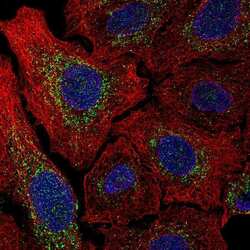
- Immunofluorescent staining of MCU in human A549 cells using the MCU monoclonal antibody (Product # MA5-24702), showing specific staining in the mitochondria in green. Microtubule- and nuclear probes are visualized in red and blue, respectively (where available).

Supportive validation
- Submitted by
- Invitrogen Antibodies (provider)
- Main image

- Experimental details
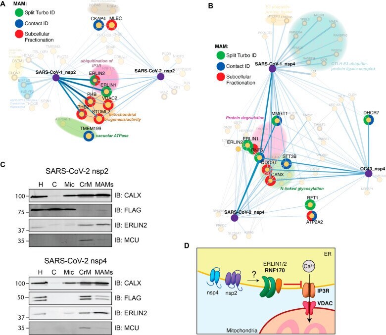
- Figure 5 Enrichment of MAM proteins as nsp2 and nsp4 interactors. (A, B) Interactors of nsp2 (A) and nsp4 (B) homologue annotated for MAM proteins. The lists of interactors was cross referenced with previous publications profiling the MAM proteome (Split-Turbo ID, Contact-ID, and subcellular fractionation ). (C) Subcellular fractions of SARS-CoV-2 nsp2 or nsp4 transfected HEK293T cells to determine the localization of viral proteins to MAMs. Homogenate (H), cytosol (C), microsome (Mic), crude mitochondria (CrM), and MAMs fractions were probed via western blot for subcellular markers (CALX and ERLIN2 for MAMs; MCU for mitochondria) and viral proteins (FLAG). Subcellular fractionation was performed in triplicate, and representative blots are shown. (D) Proposed model for how SARS-CoV nsp2 and nsp4 utilize ERLIN1/2 and interacting protein factors to regulate ER Ca 2+ signaling at MAMs.