Explore
 Validate
 Learn
Learn Western blot
 ELISA
ELISA Immunocytochemistry
Antibody data
- Antibody Data
- Antigen structure
- References [1]
- Comments [0]
- Validations
- Immunocytochemistry [5]
- Other assay [1]
Submit
Validation data
Reference
Comment
Report error
- Product number
- PA5-120437 - Provider product page

- Provider
- Invitrogen Antibodies
- Product name
- MCU Polyclonal Antibody
- Antibody type
- Polyclonal
- Antigen
- Recombinant protein fragment
- Description
- Positive test controls include: HepG2. Immunogen sequence: VHQRIASWQN LGAVYCSTVV PSDDVTVVYQ NGLPVISVRL PSRRERCQFT LKPISDSVGV FLRQLQEEDR GIDRVAIYSP DGVRVAASTG IDLLLLDDFK LVINDLTYHV RPPKRDLLSH
- Reactivity
- Human, Mouse, Rat
- Host
- Rabbit
- Isotype
- IgG
- Vial size
- 100 μL
- Concentration
- 2.07 mg/mL
- Storage
- -20°C, Avoid Freeze/Thaw Cycles
Submitted references Effect of ginkgolide K on calcium channel activity in Alzheimer's disease.
Liu H, Li Q, Zhang X, Shi Y, Li J
Experimental and therapeutic medicine 2022 Jun;23(6):426
Experimental and therapeutic medicine 2022 Jun;23(6):426
No comments: Submit comment
Supportive validation
- Submitted by
- Invitrogen Antibodies (provider)
- Main image
- Experimental details
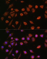
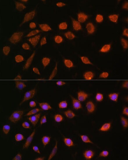
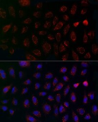
- Immunocytochemical analysis of MCU in L929 cells using a MCU Polyclonal antibody (Product # PA5-120437). Blue: DAPI for nuclear staining.
- Submitted by
- Invitrogen Antibodies (provider)
- Main image
- Experimental details
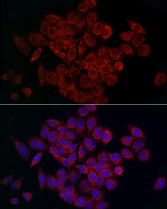
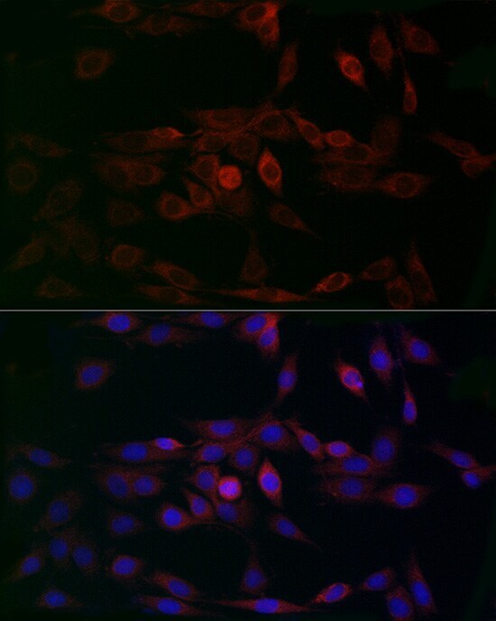
- Immunofluorescence analysis of MCU in A-549 cells. Samples were incubated with MCU Polyclonal antibody (Product # PA5-120437) using a dilution of 1:200 (40x lens). Blue: DAPI for nuclear staining.
- Submitted by
- Invitrogen Antibodies (provider)
- Main image

- Experimental details
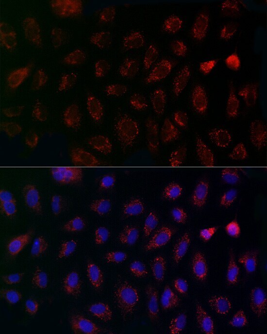
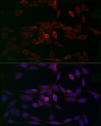
- Immunofluorescence analysis of MCU in HeLa cells. Samples were incubated with MCU Polyclonal antibody (Product # PA5-120437) using a dilution of 1:200 (40x lens). Blue: DAPI for nuclear staining.
- Submitted by
- Invitrogen Antibodies (provider)
- Main image
- Experimental details
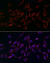
- Immunofluorescence analysis of MCU in NIH/3T3 cells. Samples were incubated with MCU Polyclonal antibody (Product # PA5-120437) using a dilution of 1:200 (40x lens). Blue: DAPI for nuclear staining.
- Submitted by
- Invitrogen Antibodies (provider)
- Main image
- Experimental details
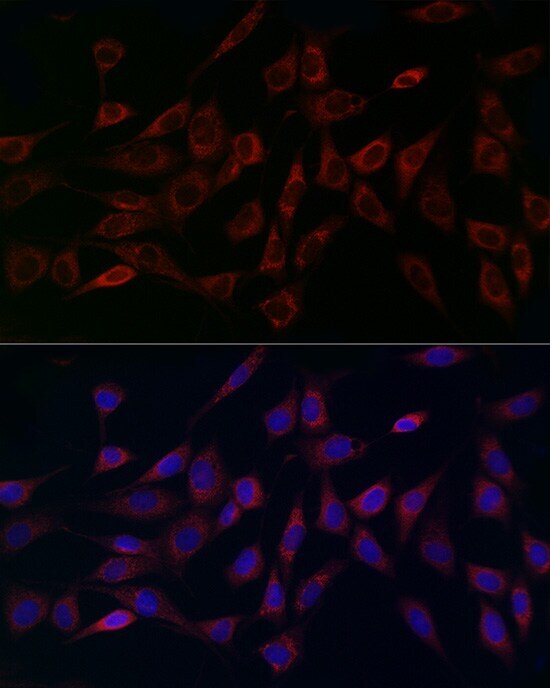
- Immunofluorescence analysis of MCU in PC-12 cells. Samples were incubated with MCU Polyclonal antibody (Product # PA5-120437) using a dilution of 1:200 (40x lens). Blue: DAPI for nuclear staining.
Supportive validation
- Submitted by
- Invitrogen Antibodies (provider)
- Main image
- Experimental details
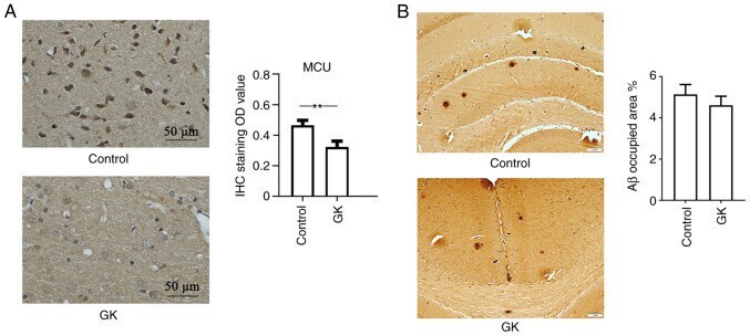
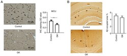
- Figure 7 MCU expression and Abeta deposits in APP/PS1 mice with GK supplementation. APP/PS1 mice (age, 6 months old; n=10 mice/group; male-to-female ratio, 1:1) were administered 8 mg/kg GK intraperitoneally for 1 month, while mice treated with DMSO were used as controls. After the treatments, the mouse brains were collected and cut into sections. The sections were then stained with antibody against MCU. The staining results were visualized using a light microscope. (A) IHC staining of the cortex of mice indicated clear positive expression of MCU in neuronal cells. Quantitative data indicated that GK supplementation significantly inhibited the expression of MCU in the cerebral cortex of mice. The expression levels of MCU were semiquantified according to the standard IHC staining grade system (scale bar, 50 um). (B) IHC staining of brain tissues was performed using antibodies against Abeta. Abeta load was estimated by stereology and presented as % positive staining of the image area. Qualitative assessment of Abeta deposition did not indicate any significant difference in the number of Abeta plaques between mice with and those without GK administration (scale bar, 1 mm). Values are expressed as the mean +- standard error of the mean. ** P