Explore
 Validate
 Learn
Learn Western blot
Antibody data
- Antibody Data
- Antigen structure
- References [0]
- Comments [0]
- Validations
- Western blot [2]
- Immunohistochemistry [1]
Submit
Validation data
Reference
Comment
Report error
- Product number
- AGC-040-50UL - Provider product page

- Provider
- Invitrogen Antibodies
- Product name
- GRIK3 (GluK3) (extracellular) Polyclonal Antibody
- Antibody type
- Polyclonal
- Antigen
- Other
- Reactivity
- Human, Mouse, Rat
- Host
- Rabbit
- Isotype
- IgG
- Vial size
- 50 µL
- Concentration
- 0.8 mg/mL
- Storage
- -20° C, Avoid Freeze/Thaw Cycles
No comments: Submit comment
Supportive validation
- Submitted by
- Invitrogen Antibodies (provider)
- Main image

- Experimental details
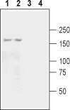
- Western blot analysis of mouse (lanes 1 and 3) and rat (lanes 2 and 4) brain membranes: - 1,2. Anti-GRIK3 (GluK3) (extracellular) Antibody (#AGC-040), (1:200).3,4. Anti-GRIK3 (GluK3) (extracellular) Antibody , preincubated with GRIK3/GluK3 (extracellular) Blocking Peptide (#BLP-GC040).
- Submitted by
- Invitrogen Antibodies (provider)
- Main image
- Experimental details
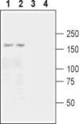
- Western blot analysis of mouse (lanes 1 and 3) and rat (lanes 2 and 4) brain membranes: - 1,2. Anti-GRIK3 (GluK3) (extracellular) Antibody (#AGC-040), (1:200).3,4. Anti-GRIK3 (GluK3) (extracellular) Antibody , preincubated with GRIK3/GluK3 (extracellular) Blocking Peptide (#BLP-GC040).

Supportive validation
- Submitted by
- Invitrogen Antibodies (provider)
- Main image
- Experimental details
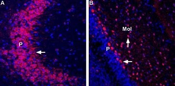
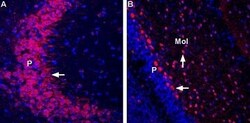
- Expression of GluR7 in mouse hippocampus and rat cerebellum - Immunohistochemical staining of immersion-fixed, free floating frozen brain sections using Anti-GRIK3 (GluK3) (extracellular) Antibody (#AGC-040), (1:400).A. GluR7 staining (red) in mouse hippocampus reveals expression in the pyramidal (P) layer (arrow).B. GluR7 staining (red) in rat cerebellum reveals expression in the Purkinje (P) layer (horizontal arrow) and in the molecular layer (MOL) interneurons (arrow).