 Explore
 Validate
 Learn
Learn Western blot
 Immunocytochemistry
Antibody data
- Antibody Data
- Antigen structure
- References [1]
- Comments [0]
- Validations
- Immunocytochemistry [3]
- Immunohistochemistry [1]
- Other assay [2]
Submit
Validation data
Reference
Comment
Report error
- Product number
- PA5-21418 - Provider product page

- Provider
- Invitrogen Antibodies
- Product name
- NULP1 Polyclonal Antibody
- Antibody type
- Polyclonal
- Antigen
- Recombinant full-length protein
- Description
- Recommended positive controls: 293T, HeLa, Molt-4, mouse brain. Predicted reactivity: Mouse (93%), Bovine (90%). Store product as a concentrated solution. Centrifuge briefly prior to opening the vial.
- Reactivity
- Human, Mouse
- Host
- Rabbit
- Isotype
- IgG
- Vial size
- 100 μL
- Concentration
- 1 mg/mL
- Storage
- Store at 4°C short term. For long term storage, store at -20°C, avoiding freeze/thaw cycles.
Submitted references Identification and characterization of distinct brown adipocyte subtypes in C57BL/6J mice.
Karlina R, Lutter D, Miok V, Fischer D, Altun I, Schöttl T, Schorpp K, Israel A, Cero C, Johnson JW, Kapser-Fischer I, Böttcher A, Keipert S, Feuchtinger A, Graf E, Strom T, Walch A, Lickert H, Walzthoeni T, Heinig M, Theis FJ, García-Cáceres C, Cypess AM, Ussar S
Life science alliance 2021 Jan;4(1)
Life science alliance 2021 Jan;4(1)
No comments: Submit comment
Supportive validation
- Submitted by
- Invitrogen Antibodies (provider)
- Main image
- Experimental details
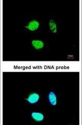
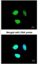
- Immunofluorescent analysis of NULP1 in paraformaldehyde-fixed HeLa cells using a NULP1 polyclonal antibody (Product # PA5-21418) at a 1:200 dilution.
- Submitted by
- Invitrogen Antibodies (provider)
- Main image
- Experimental details
- Immunofluorescence analysis of paraformaldehyde-fixed HeLa, using NULP1 antibody (Product # PA5-21418) at 1:200 dilution.

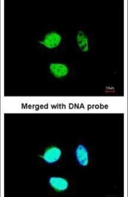
- Submitted by
- Invitrogen Antibodies (provider)
- Main image

- Experimental details
- Immunofluorescence analysis of paraformaldehyde-fixed HeLa, using NULP1 antibody (Product # PA5-21418) at 1:200 dilution.

Supportive validation
- Submitted by
- Invitrogen Antibodies (provider)
- Main image

- Experimental details
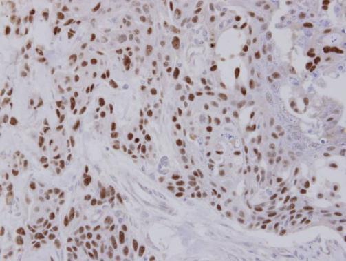
- Immunohistochemical analysis of paraffin-embedded FaDu xenograft, using NULP1 (Product # PA5-21418) antibody at 1:100 dilution. Antigen Retrieval: Citrate buffer, pH 6.0, 15 min.
Supportive validation
- Submitted by
- Invitrogen Antibodies (provider)
- Main image
- Experimental details
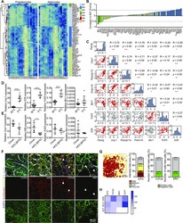
- Figure 6. EIF5, TCF25, and BIN1 mark subsets of brown adipocytes. (A) Heat maps of stably expressed genes of preadipocytes (left panel) and differentiated brown adipocytes (right panel). Module membership is indicated by the right color bar. (B) Correlation coefficients for stable expressed genes compared with estimated brown adipose tissue (BAT)ness. Color of bars indicates module membership. (C) Correlation plot for pairwise comparison of selected markers to selected stably expressed genes. Red plots denote a significant correlation. (D) Expression of Ucp1 , Eif5 , Tcf25 , and Bin1 in mice kept in cold or thermoneutrality (n = 6-8, data are mean expressions normalized to B2m +- SEM). (E) Expression analysis of Ucp1 , Eif5 , Tcf25 , and Bin1 in chow- and high-fat diet-fed mice (n = 4-6, data were mean expressions normalized to B2m +- SEM). (F) BAT co-staining for EIF5, TCF25 or BIN1 (red) and UCP1 (green), with F-actin (gray) and Dapi (blue) from wild-type C57BL/6J mice. Arrows indicate nuclear staining. (G) Representative color gradient picture of EIF5 staining on BAT (left panel), and percentage area of different EIF5 intensity (high, medium, low, and very low) normalized to total area (second panel, n = 8 sections). Quantification of TCF25- and BIN1-positive and negative cells in percentage of total cells (n = 9-18 sections). (H) Heat map of mRNA expression in human periadrenal (ADR), supraclavicular (SCLV), and subcutaneous (SCT) adipose tissue from six different donors
- Submitted by
- Invitrogen Antibodies (provider)
- Main image
- Experimental details
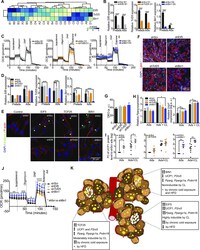
- Figure 7. Loss of Bin1 increases basal respiration and UCP1 protein content. (A) Heat map and hierarchical clustering of cell lines with regard to the mRNA expressions of Eif5 , Tcf25 and Bin1 . (B) Expression of Eif5 , Tcf25 , and Bin1 in control cells (shScr) of different clones (B1, D1, and D5) and the respective knockdown (shEif5 B1, shTcf25 D1, and shBin1 D5); Preads (d0 of differentiation) and Ads (d8 of differentiation) (n = 5). (C) Oxygen consumption rate of shScr B1 and shEif5 B1 (left panel), shScr D1, and shTcf25 D1 (middle panel) and shScr D5 and shBin1 D5 (right panel) at day 8 of differentiation measured by Seahorse (n = 4-5). (D) Expression of Eif5 , Tcf25 , and Bin1 in control cells (shScr) and respective knock-down (shEif5, shTcf25, and shBin1); Preads (d0 of differentiation) and Ads (d8 of differentiation) (n = 5). (E) Immunofluorescence staining of EIF5/TCF25/BIN1 (green), F-actin (red), and DAPI (blue) on shScr/shEif5/shTcf25/shBin1 preadipocytes. (F) Representative images of differentiated cell lines (shScr, shEif5, shTcf25, and shBin1) stained with F-actin (white), lipid droplets (red), and DAPI (blue). (G) Quantification of relative lipid accumulation measured by Oil Red O staining at day 8 of differentiation from knockdown cell lines (n = 4, mean OD normalized to DAPI +- SEM). (H) mRNA expressions of Pparg (left panel) and Ucp1 (right panel) on shScr, shEif5, shTcf25, and shBin1 adipocytes and CL-316,243-treated adipocytes (0.5 muM CL-316,243 for 3 h;
