Explore
 Validate
 Learn
Learn Western blot
 Immunocytochemistry
Antibody data
- Antibody Data
- Antigen structure
- References [0]
- Comments [0]
- Validations
- Immunocytochemistry [3]
- Immunoprecipitation [1]
- Immunohistochemistry [7]
- Other assay [1]
Submit
Validation data
Reference
Comment
Report error
- Product number
- PA5-63081 - Provider product page

- Provider
- Invitrogen Antibodies
- Product name
- VGF Polyclonal Antibody
- Antibody type
- Polyclonal
- Antigen
- Recombinant protein fragment
- Description
- Immunogen sequence: APARDELPDW NEVLPPWDRE EDEVYPPGPY HPFPNYIRPR TLQPPSALRR RHYHHALPPS RHYPGREAQA RRAQEEAEAE ERRLQEQEEL ENYIEHV Highest antigen sequence identity to the following orthologs: Mouse - 90%, Rat - 89%.
- Reactivity
- Human
- Host
- Rabbit
- Isotype
- IgG
- Vial size
- 100 μL
- Concentration
- 0.10 mg/mL
- Storage
- Store at 4°C short term. For long term storage, store at -20°C, avoiding freeze/thaw cycles.
No comments: Submit comment
Supportive validation
- Submitted by
- Invitrogen Antibodies (provider)
- Main image
- Experimental details
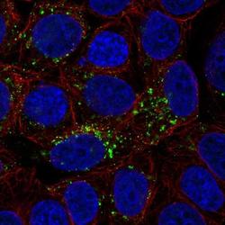
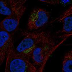
- Immunofluorescent staining of VGF in human cell line MCF7 shows positivity in vesicles. Samples were probed using a VGF Polyclonal Antibody (Product # PA5-63081).
- Submitted by
- Invitrogen Antibodies (provider)
- Main image
- Experimental details
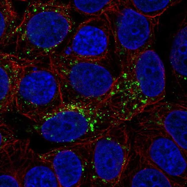
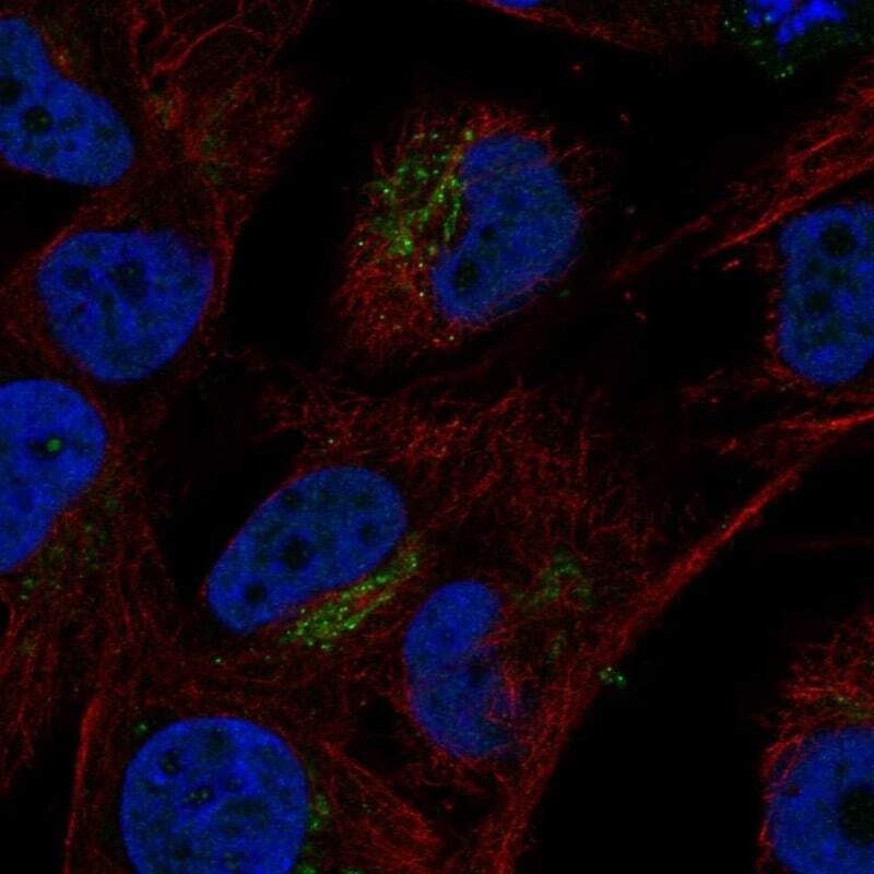
- Immunofluorescent staining of VGF in human cell line SK-MEL-30 using VGF Polyclonal Antibody (Product # PA5-63081) shows localization to the Golgi apparatus.

- Submitted by
- Invitrogen Antibodies (provider)
- Main image
- Experimental details
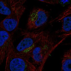
- Immunofluorescent staining of VGF in human cell line SK-MEL-30 using VGF Polyclonal Antibody (Product # PA5-63081) shows localization to the Golgi apparatus.
Supportive validation
- Submitted by
- Invitrogen Antibodies (provider)
- Main image
- Experimental details
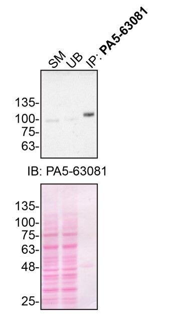
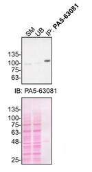
- U2OS cells were washed 3x with PBS and starved for ~18 hrs in DMEM high glucose containing L-glutamate and penicillin/streptomycin. Immunoprecipitation of VGF was performed on U2OS concentrated cell culture media, concentrated by centrifuging for 10 min at 500 x g to eliminate cells and larger contaminants, then for 10 min at 4500 x g to eliminate smaller contaminants. Culture media were concentrated using centrifugal filter units at 4000 x g for 15 min. The resulting 500 μl of the concentrated media were centrifuged again at 4000 x g for 15 min using centrifugal filter units with a membrane NMWL of 50 kDa. Antibody-bead conjugates were prepared by adding 1 µg of VGF polyclonal antibody (Product # PA5-63081) with 30 µL of protein A-Sepharose beads and rocked overnight at 4°C. 0.85 mg of lysate was incubated with an antibody-bead conjugate for 2 hours at 4°C. Following centrifugation and multiple washes, 10% starting material (SM), 10% unbound fraction (UB) and immunoprecipitated fraction (IP) were processed for immunoblot using the same VGF polyclonal antibody. Ponceau stained transfer of blot is shown. Data courtesy of YCharOS Inc., an open science company with the mission of characterizing commercially available antibodies using knockout validation.

Supportive validation
- Submitted by
- Invitrogen Antibodies (provider)
- Main image
- Experimental details
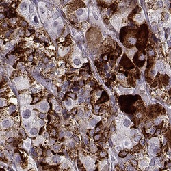
- Immunohistochemical staining of VGF in human pituitary gland using VGF Polyclonal Antibody (Product # PA5-63081).
- Submitted by
- Invitrogen Antibodies (provider)
- Main image

- Experimental details
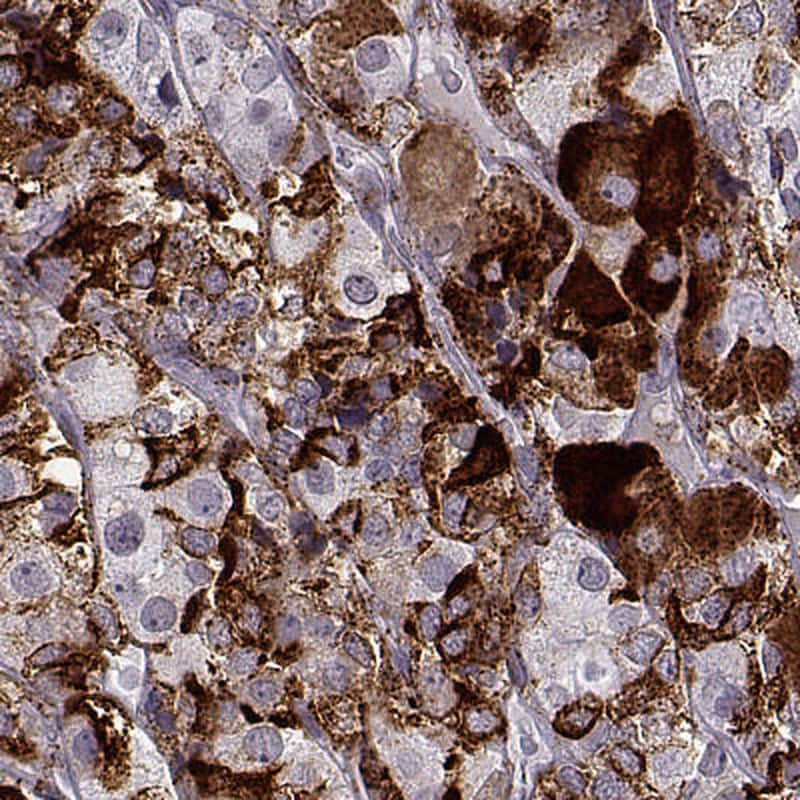
- Immunohistochemical staining of VGF in human pituitary gland using a VGF Polyclonal Antibody (Product # PA5-63081) shows strong cytoplasmic positivity in anterior cells.

- Submitted by
- Invitrogen Antibodies (provider)
- Main image

- Experimental details
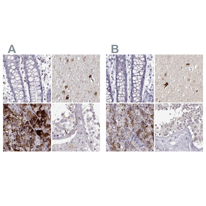
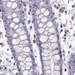
- Immunohistochemical staining of VGF in human colon, hypothalamus, pituitary gland and testis using VGF Polyclonal Antibody (Product # PA5-63081) (A) shows similar protein distribution across tissues to an independent VGF Polyclonal Antibody (B).
- Submitted by
- Invitrogen Antibodies (provider)
- Main image
- Experimental details
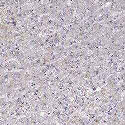
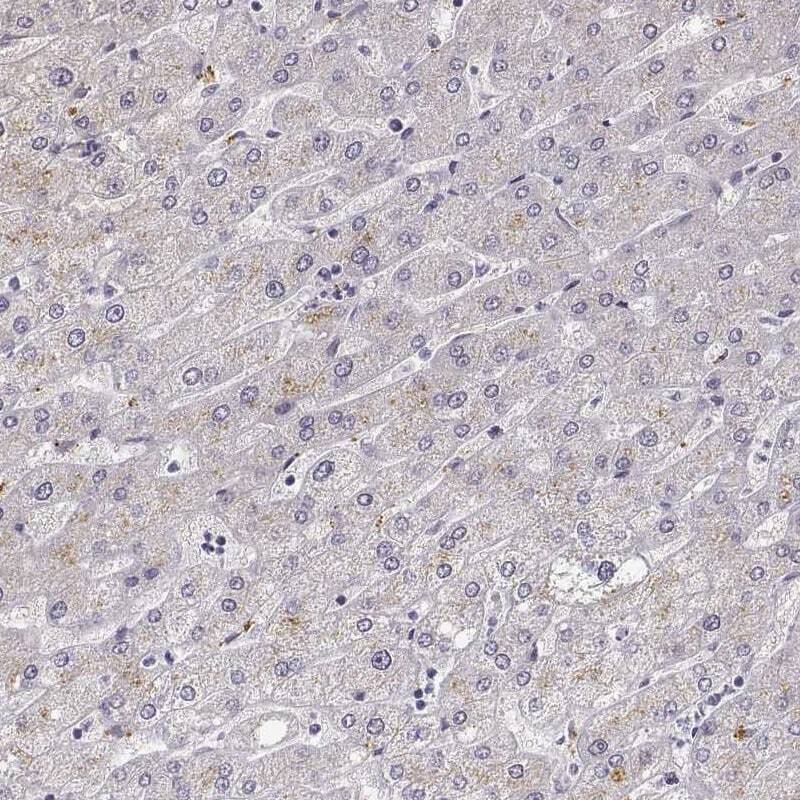
- Immunohistochemical staining of VGF in human liver using VGF Polyclonal Antibody (Product # PA5-63081) shows low expression as expected.
- Submitted by
- Invitrogen Antibodies (provider)
- Main image
- Experimental details
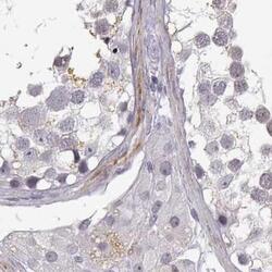
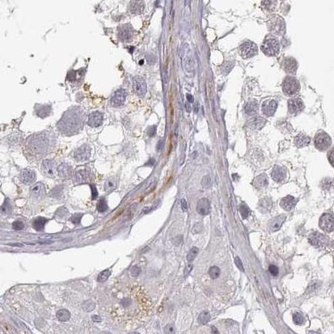
- Immunohistochemical staining of VGF in human testis using VGF Polyclonal Antibody (Product # PA5-63081).
- Submitted by
- Invitrogen Antibodies (provider)
- Main image
- Experimental details
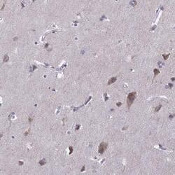
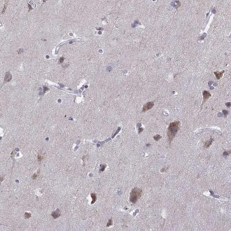
- Immunohistochemical staining of VGF in human cerebral cortex using VGF Polyclonal Antibody (Product # PA5-63081) shows high expression.
- Submitted by
- Invitrogen Antibodies (provider)
- Main image
- Experimental details
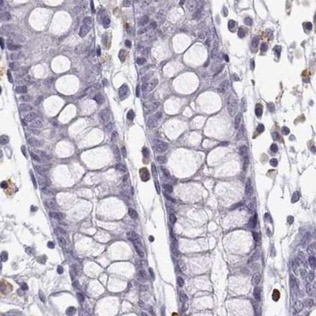
- Immunohistochemical staining of VGF in human colon using VGF Polyclonal Antibody (Product # PA5-63081).
Supportive validation
- Submitted by
- Invitrogen Antibodies (provider)
- Main image

- Experimental details
- U2OS cells were washed 3x with PBS and starved for ~18 hrs in DMEM high glucose containing L-glutamate and penicillin/streptomycin. Immunoprecipitation of VGF was performed on U2OS concentrated cell culture media, concentrated by centrifuging for 10 min at 500 x g to eliminate cells and larger contaminants, then for 10 min at 4500 x g to eliminate smaller contaminants. Culture media were concentrated using centrifugal filter units at 4000 x g for 15 min. The resulting 500 μl of the concentrated media were centrifuged again at 4000 x g for 15 min using centrifugal filter units with a membrane NMWL of 50 kDa. Antibody-bead conjugates were prepared by adding 1 µg of VGF polyclonal antibody (Product # PA5-63081) with 30 µL of protein A-Sepharose beads and rocked overnight at 4°C. 0.85 mg of lysate was incubated with an antibody-bead conjugate for 2 hours at 4°C. Following centrifugation and multiple washes, 10% starting material (SM), 10% unbound fraction (UB) and immunoprecipitated fraction (IP) were processed for immunoblot using the same VGF polyclonal antibody. Ponceau stained transfer of blot is shown. Data courtesy of YCharOS Inc., an open science company with the mission of characterizing commercially available antibodies using knockout validation.