Explore
 Validate
 Learn
Learn Western blot
Antibody data
- Antibody Data
- Antigen structure
- References [0]
- Comments [0]
- Validations
- Western blot [1]
- Immunocytochemistry [1]
- Immunohistochemistry [1]
Submit
Validation data
Reference
Comment
Report error
- Product number
- AP11318PU-N - Provider product page

- Provider
- Acris Antibodies GmbH
- Proper citation
- Acris Antibodies GmbH Cat#AP11318PU-N, RRID:AB_1770064
- Product name
- anti NIP3 (BH3 Domain)
- Antibody type
- Polyclonal
- Antigen
- This antibody is generated from rabbits immunized with a KLH conjugated synthetic peptide selected from the N-terminal region (BH3 domain) of human NIP3.
- Reactivity
- Human, Mouse
- Host
- Rabbit
- Vial size
- 0.4 ml
- Concentration
- lot specific
No comments: Submit comment
Supportive validation
- Submitted by
- Acris Antibodies GmbH (provider)
- Main image
- Experimental details
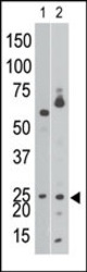
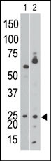
- Western blot analysis using anti-NIP3 BH3 domain Pab (Cat. #AP11318PU-N) to detect NIP3 BH3 in Ramos cell lysate (lane 1) and in mouse brain tissue lysate (lane 2).
Supportive validation
- Submitted by
- Acris Antibodies GmbH (provider)
- Main image
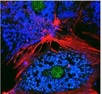
- Experimental details
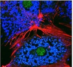
- Freshly isolated mouse hepatocytes plated on coverslips (2 x10e5 cells/22-mm glass coverslip) were cultured under normoxic conditions for 6 hr. The cells were then fixed in 2% paraformaldehyde in PBS for 1 hr, and processed for confocal immunofluorescence (red: F-actin, blue: ATP-synthase, green: BNIP3). Fluorescence labeling of BNIP3 accomplished with anti-BNIP3 antibody (Cat. #AP11318PU-N). Data courtesy of Ruben Zamora, University of Pittsburgh.
Supportive validation
- Submitted by
- Acris Antibodies GmbH (provider)
- Main image
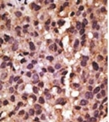
- Experimental details
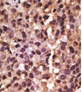
- Formalin-fixed and paraffin-embedded human cancer tissue reacted with the primary antibody, which was peroxidase-conjugated to the secondary antibody, followed by DAB staining. This data demonstrates the use of this antibody for immunohistochemistry; clinical relevance has not been evaluated.